
Page 384 - Feline diagnostic imaging
P. 384
23.6 Abnormal AApmombnp nof tpf pallbp lipo 393
(a) (b)
(c) (d)
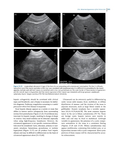
Figure 23.25 Longitudinal ultrasound image of the liver of a cat presenting with anorexia and constipation. The liver is diffusely
hypoechoic (a,b). Fine needle aspiration of the liver was consistent with lymphosarcoma. In a different cat presenting for decreased
appetite, multiple well-defined masses are identified within the liver parenchyma (c). The outer border of these masses is hyperechoic
and the central region is hypoechoic (d). Fine needle aspiration of the masses was consistent with large granular lymphocytic
lymphoma. Source: Images courtesy of Dr Merrilee Holland, Auburn University.
hepatic echogenicity should be correlated with clinical Ultrasound can be extremely useful in differentiating
signs and bloodwork, and a biopsy is necessary for defini- cystic versus solid masses; focal, multifocal, or diffuse
tive diagnosis. Prebiopsy coagulation screening is a useful distribution of masses; and the relation of the mass to
precaution when liver disease is suspected. adjacent structures, such as large blood vessels or the
Focal hepatic disease appears as a nodule or mass that gallbladder. Hepatic neoplasia has a variable appear-
differs in texture and/or echogenicity from surrounding ance [42,51–53,55,66,69,70]. The most common hepatic
normal liver parenchyma (Figure 23.26). Focal disease may tumor in the cat is biliary cystadenoma [70–76]. These
interrupt the hepatic margin, resulting in change of shape are benign cystic hepatic tumors seen mainly in
or contour. Very small nodules can be detected, especially older cats and may be focal or multifocal. Although
when using high‐frequency transducers. However, the variable in appearance, the presence of a cystic compo -
ultrasound appearance is not specific; numerous differen- nent somewhere in the mass is a consistent finding
tials must be considered. Cysts, abscesses, primary or met- (Figures 23.28 and 23.29). Biliary cystadenomas may
astatic neoplasia, hematomas, granulomas, or nodular appear multilocular, containing thin‐walled cysts, or as
hyperplasia (Figure 23.27) can all produce focal hepatic hyperechoic masses with a cystic component. Most cystic
disease and may be difficult to differentiate on the basis of portions of these masses will be characterized by acous-
ultrasound appearance alone [51–53,68]. tic enhancement.