
Page 387 - Feline diagnostic imaging
P. 387
396 23 Liver
sometimes dilated bile duct (Figures 23.22 and 23.37–
23.39) [18,55,60,61,86,87]. The liver may be normal in size
or enlarged, with a hypoechoic, heterogeneous, or hypere-
choic (likely due to hepatic lipidosis) parenchyma
(Figures 23.18 and 23.19). Pancreatitis and inflammatory
bowel disease are often present concurrently (triaditis),
and may occur due to the unique feline biliary anatomy,
where the bile duct and pancreatic duct enter the duode-
num together at the major duodenal papilla (Figure 23.40)
[60,61,85,86,88–93].
Other causes of gallbladder wall thickening include
edema with a thickened hypoechoic wall with echogenic
inner and outer rims, creating a layered appearance. This
can occur secondary to right‐sided congestive heart failure,
hypoalbuminemia, sepsis, anaphylactic reaction, and neo-
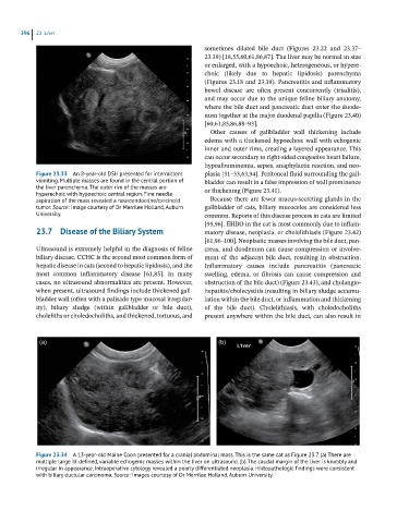
Figure 23.33 An 8-year-old DSH presented for intermittent plasia [51–53,63,94]. Peritoneal fluid surrounding the gall-
vomiting. Multiple masses are found in the central portion of bladder can result in a false impression of wall prominence
the liver parenchyma. The outer rim of the masses are or thickening (Figure 23.41).
hyperechoic with hypoechoic central region. Fine needle
aspiration of the mass revealed a neuroendocrine/carcinoid Because there are fewer mucus‐secreting glands in the
tumor. Source: Image courtesy of Dr Merrilee Holland, Auburn gallbladder of cats, biliary mucoceles are considered less
University. common. Reports of this disease process in cats are limited
[95,96]. EHBO in the cat is most commonly due to inflam-
23.7 Disease of the Biliary System matory disease, neoplasia, or cholelithiasis (Figure 23.42)
[61,96–100]. Neoplastic masses involving the bile duct, pan-
Ultrasound is extremely helpful in the diagnosis of feline creas, and duodenum can cause compression or involve-
biliary disease. CCHC is the second most common form of ment of the adjacent bile duct, resulting in obstruction.
hepatic disease in cats (second to hepatic lipidosis), and the Inflammatory causes include pancreatitis (pancreatic
most common inflammatory disease [61,85]. In many swelling, edema, or fibrosis can cause compression and
cases, no ultrasound abnormalities are present. However, obstruction of the bile duct) (Figure 23.43), and cholangio-
when present, ultrasound findings include thickened gall- hepatitis/cholecystitis (resulting in biliary sludge accumu-
bladder wall (often with a palisade type mucosal irregular- lation within the bile duct, or inflammation and thickening
ity), biliary sludge (within gallbladder or bile duct), of the bile duct). Cholelithiasis, with choledocholiths
choleliths or choledocholiths, and thickened, tortuous, and present anywhere within the bile duct, can also result in
(a) (b)
Figure 23.34 A 13-year-old Maine Coon presented for a cranial abdominal mass. This is the same cat as Figure 23.7. (a) There are
multiple large ill-defined, variable echogenic masses within the liver on ultrasound. (b) The caudal margin of the liver is knobbly and
irregular in appearance. Intraoperative cytology revealed a poorly differentiated neoplasia. Histopathologic findings were consistent
with biliary ductular carcinoma. Source: Images courtesy of Dr Merrilee Holland, Auburn University.