
Page 390 - Feline diagnostic imaging
P. 390
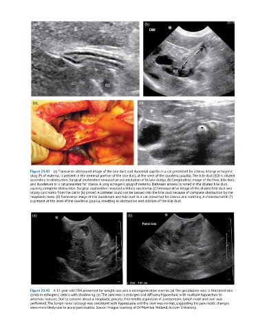
(a)                                                  (b)


                 (c)                                               (d)


               Figure 23.42  (a) Transverse ultrasound image of the bile duct and duodenal papilla in a cat presented for icterus. A large echogenic
               plug (P) of material is present in the terminal portion of the bile duct, at the level of the duodenal papilla. The bile duct (BD) is dilated
               secondary to obstruction. Surgical exploration revealed an accumulation of biliary sludge. (b) Longitudinal image of the liver, bile duct,
               and duodenum in a cat presented for icterus. A long echogenic plug of material (between arrows) is noted in the dilated bile duct,
               causing complete obstruction. Surgical exploration revealed a biliary carcinoma. (c) Intraoperative image of the dilated bile duct and
               biliary carcinoma from the cat in (b) (arrow). A catheter could not be passed into the bile duct because of complete obstruction by the
               neoplastic mass. (d) Transverse image of the duodenum and bile duct in a cat presented for icterus and vomiting. A choledocholith (*)
               is present at the level of the duodenal papilla, resulting in obstruction and dilation of the bile duct.


                (a)                                           (b)


               Figure 23.43  A 12-year-old DSH presented for weight loss and a nonregenerative anemia. (a) The gallbladder wall is thickened and
               contains echogenic debris with shadowing. (b) The pancreas is enlarged and diffusely hypoechoic with multiple hypoechoic to
               anechoic nodules. Due to concern about a neoplastic process, fine needle aspiration of a mesenteric lymph node and liver was
               performed. The lymph node cytology was consistent with hyperplasia and the liver was normal, suggesting the pancreatic changes
               were more likely due to acute pancreatitis. Source: Images courtesy of Dr Merrilee Holland, Auburn University.