
Page 388 - Feline diagnostic imaging
P. 388
(a) (b)
(c)
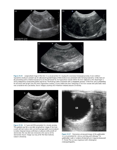
Figure 23.35 Longitudinal image of the liver in a cat presented for staging for cutaneous hemangiosarcoma. A well-defined
hypoechoic nodule is present (a). An ultrasound-guided biopsy of this nodule was consistent with hemangiosarcoma. In the next cat
(b) presenting with a rhabdomyosarcoma on the left forelimb, the hypoechoic nodule within the liver adjacent to the diaphragm is
being sampled by ultrasound-guided aspiration. The findings were consistent with a neoplastic process. In the final cat (c) presenting
for anorexia and weight loss, the small hypoechoic nodule is found within the liver. Sampling of the liver nodule and pancreatic mass
was consistent with carcinoma. Source: Images courtesy of Dr Merrilee Holland, Auburn University.
Figure 23.36 A 7-year-old DSH presented for chronic anemia.
This patient was fed a raw diet. Longitudinal image of the liver;
a well-defined nodule with central focal gas (with reverberation)
is noted. Surgical biopsy confirmed an abscess with growth of
Salmonella. The raw diet was believed to the source of the
Salmonella. Source: Image courtesy of Dr Merrilee Holland, Figure 23.37 Transverse ultrasound image of the gallbladder
Auburn University. in a cat presented for icterus, vomiting, and lethargy. The
gallbladder wall is markedly thickened and irregular (measured
2–3 mm). The cat was diagnosed with cholangitis/
cholangiohepatitis.