
Page 386 - Feline diagnostic imaging
P. 386
23.6 Abnormal AApmombnp nof tpf pallbp lipo 395
Figure 23.29 A 2-year-old domestic shorthair (DSH) presented
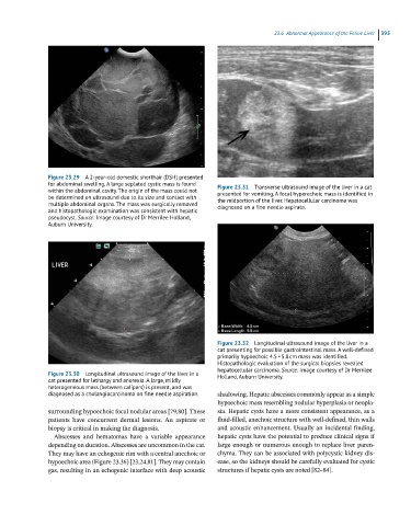
for abdominal swelling. A large septated cystic mass is found Figure 23.31 Transverse ultrasound image of the liver in a cat
within the abdominal cavity. The origin of the mass could not presented for vomiting. A focal hyperechoic mass is identified in
be determined on ultrasound due to its size and contact with the midportion of the liver. Hepatocellular carcinoma was
multiple abdominal organs. The mass was surgically removed diagnosed on a fine needle aspirate.
and histopathologic examination was consistent with hepatic
pseudocyst. Source: Image courtesy of Dr Merrilee Holland,
Auburn University.
Figure 23.32 Longitudinal ultrasound image of the liver in a
cat presenting for possible gastrointestinal mass. A well-defined
primarily hypoechoic 4.5 × 5.8 cm mass was identified.
Histopathologic evaluation of the surgical biopsies revealed
hepatocellular carcinoma. Source: Image courtesy of Dr Merrilee
Figure 23.30 Longitudinal ultrasound image of the liver in a Holland, Auburn University.
cat presented for lethargy and anorexia. A large, mildly
heterogeneous mass (between calipers) is present, and was
diagnosed as a cholangiocarcinoma on fine needle aspiration. shadowing. Hepatic abscesses commonly appear as a simple
hypoechoic mass resembling nodular hyperplasia or neopla-
surrounding hypoechoic focal nodular areas [79,80]. These sia. Hepatic cysts have a more consistent appearance, as a
patients have concurrent dermal lesions. An aspirate or fluid‐filled, anechoic structure with well‐defined, thin walls
biopsy is critical in making the diagnosis. and acoustic enhancement. Usually an incidental finding,
Abscesses and hematomas have a variable appearance hepatic cysts have the potential to produce clinical signs if
depending on duration. Abscesses are uncommon in the cat. large enough or numerous enough to replace liver paren-
They may have an echogenic rim with a central anechoic or chyma. They can be associated with polycystic kidney dis-
hypoechoic area (Figure 23.36) [23,24,81]. They may contain ease, so the kidneys should be carefully evaluated for cystic
gas, resulting in an echogenic interface with deep acoustic structures if hepatic cysts are noted [82–84].