
Page 381 - Feline diagnostic imaging
P. 381
390 23 Liver
echogenic material without acoustic shadowing) is seen. 23.6 Abnormal Appearance
There is some controversy regarding the significance of of the Feline Liver
biliary sludge in dogs [45,46]. One study in cats found
that gallbladder sludge was uncommon, but was a pre - Ultrasound is helpful in differentiating between diffuse
dictive of increased liver enzymes [47]. The normal feline and focal hepatic disease. Diffuse hepatic disease can result
gallbladder wall is thin and poorly visualized, with a wall in changes in shape, size, and echogenicity [51–56].
thickness of less than 1 mm or not visualized at all [48]. A hyperechoic liver parenchyma results in an abnormal
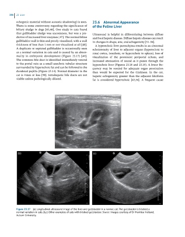
A duplicate or septated gallbladder is occasionally seen echointensity of liver to adjacent organs (hyperechoic to
as a normal variation in cats and is caused by an abnor - renal cortex, isoechoic, or hyperechoic to spleen), loss of
mality in embryonic development (Figure 23.17) [49]. visualization of the prominent periportal echoes, and
The common bile duct is identified immediately ventral increased attenuation of sound as it passes through the
to the portal vein as a small anechoic tubular structure hyperechoic liver (Figures 23.18 and 23.19). A lower fre-
surrounded by hyperechoic fat and can be followed to the quency may be needed for adequate organ penetration
duodenal papilla (Figure 23.14). Normal diameter in the than would be expected for the thickness. In the cat,
cat is 4 mm or less [50]. Intrahepatic bile ducts are not hepatic echogenicity greater than the adjacent falciform
visible unless pathologically dilated. fat is considered hyperechoic [42,56]. A frequent cause
(a)
(b) (c)
Figure 23.17 (a) Longitudinal ultrasound image of the liver and gallbladder in a normal cat. The gallbladder is bilobed, a
normal variation in cats. (b,c) Other examples of cats with bilobed gallbladder. Source: Images courtesy of Dr Merrilee Holland,
Auburn University.