
Page 377 - Feline diagnostic imaging
P. 377
386 23 Liver
(b)
(a)
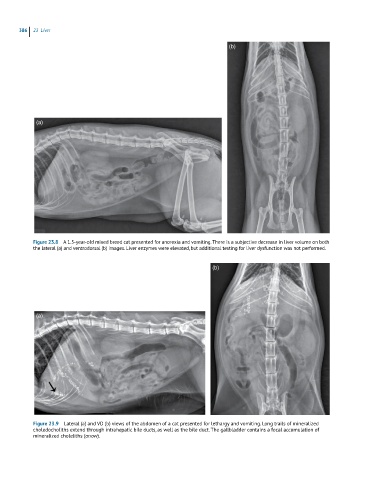
Figure 23.8 A 1.5-year-old mixed breed cat presented for anorexia and vomiting. There is a subjective decrease in liver volume on both
the lateral (a) and ventrodorsal (b) images. Liver enzymes were elevated, but additional testing for liver dysfunction was not performed.
(b)
(a)
Figure 23.9 Lateral (a) and VD (b) views of the abdomen of a cat presented for lethargy and vomiting. Long trails of mineralized
choledocholiths extend through intrahepatic bile ducts, as well as the bile duct. The gallbladder contains a focal accumulation of
mineralized choleliths (arrow).