
Page 375 - Feline diagnostic imaging
P. 375
384 23 Liver
(a)
(b)
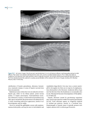
Figure 23.5 (a) Lateral image of the thorax and cranial abdomen in a cat with pleural effusion, cardiomyopathy, and pericardial
effusion. The caudal liver extends well beyond the costal arch, and has rounded, blunted margins. This is likely secondary to
congestion from elevated right heart pressure. Lateral image (b) of another cat in which disease has progressed into heart failure.
The liver extends beyond the costal arch, causing caudal deviation of the stomach (ST). Source: Images courtesy of Dr Merrilee
Holland, Auburn University.
calcification of hepatic granulomas, abscesses, hemato- nondiabetic dogs [20,21]. Cats may have a severe neutro-
mas, neoplastic masses, or areas of hepatic necrosis have philic cholangitis, but there are no reports of emphysema-
been documented. tous cholecystitis in the literature. Incidental reflux of gas
Radiolucent areas within the liver are indicative of intra- into the bile duct from the duodenum is occasionally seen
hepatic gas, either in the biliary system, portal venous in cats. This may be because of incompetence of the sphinc-
system, or hepatic parenchyma. Gastrointestinal ulcera- ter of Oddi [22].
tion, distension, trauma, or interventional procedures may Hepatic abscesses caused by gas‐forming organisms
allow gas to ascend into the portovenous circulation [4,19]. may result in gas opacities within the hepatic parenchyma
A linear, branching radiolucent appearance, similar to air [23,24]. These abscesses appear as irregularly stippled
bronchograms, may be visible. or mottled gas patterns, usually in a localized area.
Gas in or around the gallbladder occurs with emphyse- Hepatomegaly or hepatic mass is typically present with
matous cholecystitis, and may be seen in both diabetic and hepatic abscess with or without gas formation.