
Page 385 - Feline diagnostic imaging
P. 385
394 23 Liver
Figure 23.27 An ill-defined hypoechoic nodule is found in the
liver in this ultrasound image. Fine needle aspiration of the
Figure 23.26 Transverse ultrasound image of the liver in a cat nodule was consistent with a regenerative process. Source:
presented for icterus and elevated liver enzymes. A focal Image courtesy of Dr Merrilee Holland, Auburn University.
hyperechoic nodule (between calipers) is present. The cat was
diagnosed with cholangitis/cholangiohepatitis, and the etiology
of the nodule was not determined.
(a) (b) (c)
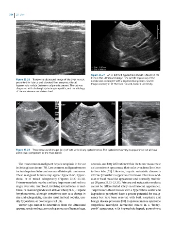
Figure 23.28 Three ultrasound images (a–c) of cats with biliary cystadenomas. The cystadenomas vary in appearance, but all have
some cystic component to the mass lesion.
The most common malignant hepatic neoplasia in the cat necrosis, and fatty infiltration within the tumor mass create
is cholangiocarcinoma [70]. Less common malignant tumors an inconsistent appearance that varies even from liver lobe
include hepatocellular carcinoma and histiocytic carcinoma. to liver lobe [55]. Likewise, hepatic metastatic disease is
These malignant tumors may appear hypoechoic, hypere- extremely variable in appearance but more often has a nod-
choic, or of mixed echogenicity (Figures 23.30–23.32). ular or focal mass‐like appearance and is usually multifo-
Primary neoplasia may be a solitary large mass confined to a cal (Figures 23.33–23.35). Primary and metastatic neoplasia
single liver lobe; multifocal, involving several lobes; or mul- cannot be differentiated solely on ultrasound appearance.
tifocal or coalescing nodules in all liver lobes [70,77]. Hepatic Target lesions (focal masses with a hyperechoic center and
lymphosarcoma, although sometimes seen as a change in hypoechoic periphery) have a greater potential for malig-
size and echogenicity, can also result in focal nodules, usu- nancy but have been reported with both neoplastic and
ally hypoechoic, or no change at all [66]. benign disease processes [78]. Hepatocutaneous syndrome
Tumor type cannot be determined from the ultrasound (superficial necrolytic dermatitis) results in a “honey-
appearance alone because varying amounts of hemorrhage, comb” appearance, with hyperechoic hepatic parenchyma