
Page 383 - Feline diagnostic imaging
P. 383
392 23 Liver
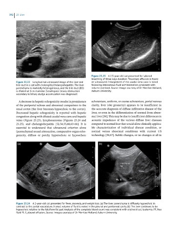
Figure 23.23 A 2.5-year-old cat presented for labored
breathing of three days duration. Tricavitary effusion is found
Figure 23.22 Longitudinal ultrasound image of the liver and on ultrasound. Enlargement of the caudal vena cava is noted
bile duct in a cat with cholangitis/cholangiohepatitis. The liver following intravenous fluid administration consistent with
parenchyma is markedly heterogeneous, and the bile duct (BD) volume overload. Source: Image courtesy of Dr Merrilee Holland,
is dilated at 1 cm diameter. Extrahepatic biliary obstruction Auburn University.
secondary to biliary sludge accumulation was diagnosed.
A decrease in hepatic echogenicity results in prominence echotexture, uniform, or coarse echotexture, portal venous
of the periportal echoes and abnormal comparison to the clarity, liver lobe geometry) appears to be insufficient in
renal cortex (the liver becomes hypoechoic to the cortex). the accurate diagnosis of diffuse infiltrative disease of the
Decreased hepatic echogenicity is reported with hepatic liver, or even in the differentiation of normal from abnor-
congestion along with dilated caudal vena cava and hepatic mal liver [58]. This may be due to insufficient differences in
veins (Figure 23.23), lymphosarcoma (Figures 23.24 and acoustic impedance of the various diffuse liver diseases
23.25), and cholangiohepatitis [52,54,55,60,63–66]. It is compared to normal liver that would allow clinically applica-
essential to understand that ultrasound criterion alone ble characterization of individual disease condition, or
(parenchymal sound attenuation, comparative organ echo- normal versus abnormal conditions with current US
genicity, diffuse or patchy hyperechoic or hypoechoic technology [58,67]. Subtle changes, or no changes at all in
(a) (b)
Figure 23.24 A 2-year-old cat presented for fever, anorexia, and weight loss. (a) The liver parenchyma is diffusely hypoechoic in
contrast to the portal vasculature. A small volume of fluid is noted in the pleural and peritoneal cavity. (b) The liver continues to be
hypoechoic relative to the falciform fat pad. Analysis of the complete blood count was consistent with erythroid cell leukemia. FF, free
fluid PL E, pleural effusion; Source: Images courtesy of Dr Merrilee Holland, Auburn University.