
Page 393 - Feline diagnostic imaging
P. 393
402 23 Liver
(a) (b)
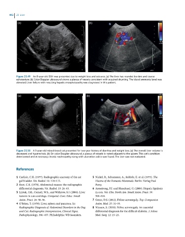
Figure 23.49 An 8-year-old DSH was presented due to weight loss and seizures. (a) The liver has rounded borders and coarse
echotexture. (b) Color Doppler ultrasound shows a plexus of vessels consistent with acquired shunting. The blood ammonia level was
elevated. Liver failure with resulting hepatic encephalopathy was diagnosed in this patient.
(a) (b)
Figure 23.50 A 9-year-old mixed-breed cat presented for two-year history of diarrhea and weight loss. (a) The overall liver volume is
decreased and hyperechoic. (b) On color Doppler ultrasound, a plexus of vessels is noted adjacent to the spleen. This cat’s condition
deteriorated and at necropsy chronic nephropathy along with ulcerative colitis was found. The liver was not evaluated.
References
1 Carlisle, C.H. (1977). Radiographic anatomy of the cat 5 Nickel, R., Schummer, A., Seiferle, E. et al. (1973). The
gallbladder. Vet. Radiol. 18: 170–172. Viscera of the Domestic Mammals. Berlin: Verlag Paul
2 Root, C.R. (1974). Abdominal masses: the radiographic Parey.
differential diagnosis. Vet. Radiol. 15: 26–43. 6 Armstrong, P.J. and Blanchard, G. (2009). Hepatic lipidosis
3 Liptak, J.M., Cernell, W.S., and Withrow, S.J. (2004). Liver in cats. Vet. Clin. North Am. Small Anim. Pract. 39:
tumors in cats and dogs. Compend. Cont. Educ. Small 599–616.
Anim. Pract. 26: 50–56. 7 Greco, D.S. (2012). Feline acromegaly. Top. Companion
4 O’Brien, T. (1978). Liver, spleen and pancreas. In: Anim. Med. 27: 31–35.
Radiographic Diagnosis of Abdominal Disorders in the Dog 8 Niessen, S. (2010). Feline acromegaly. An essential
and Cat: Radiographic Interpretation, Clinical Signs, differential diagnosis for the difficult diabetic. J. Feline
Pathophysiology, 396–457. Philadelphia: WB Saunders. Med. Surg. 12: 15–23.