
Page 392 - Feline diagnostic imaging
P. 392
23.8 Vascular Disease 401
(a) (b)
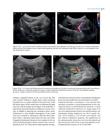
Figure 23.47 (a) A 6-month-old mixed-breed kitten presented for hypersalivation and lethargy. A small liver is noted on abdominal
ultrasound. (b) Color Doppler shows a shunt vessel bypassing the liver and heading dorsally. Partial ligation of an extrahepatic shunt
was performed at surgery.
(a) (b)
Figure 23.48 A 12-year-old DSH presented for anorexia and weight loss. The liver enzymes were elevated along with a low albumin.
On the abdominal ultrasound, a plexus of vessels is noted caudal to the left kidney seen on 2D (a) and color Doppler imaging
(b). Source: Images courtesy of Dr Merrilee Holland, Auburn University.
common congenital shunts in the cat [9,12,25–28]. The with aberrant, tortuous courses connecting the intrahepatic
most common finding is a single shunt vessel connecting portal vein and caudal vena cava and hepatic vein [105].
the portal vein, or a major tributary of the portal vein, to the Acquired shunting is uncommon in cats and has been
left lateral aspect of the caudal vena cava between the right reported in response to portal hypertension in two cats.
renal vein and the hepatic veins (Figure 23.47) [104,105]. A Acquired shunts are identified by the presence of small
right dorsal intercostal window, in addition to routine views, collateral vessels [107]. However, in a larger study of 33
may be helpful in detecting the anomalous shunt vessel. cats with splenosystemic shunts (an acquired shunt), the
Pulsed‐wave or color Doppler interrogation of the caudal underlying etiology implicated was 14/33 hepatic dis-
vena cava is helpful in assessing for abnormal flow turbu- ease, 10/33 neoplastic, 2/33 chronic renal disease, and
lence where a shunting vessel enters the vena cava [104]. five without a definitive diagnosis. The significance of
Congenital intrahepatic shunts are less common in the cat, the splenosystemic shunts has not been determined and
with left divisional (patent ductus venosus) seen most fre- they may be seen with active or previous episodes of por -
quently [9,12,25–28]. These shunt vessels typically are large, tal hypertension (Figures 23.48–23.50) [108].