
Page 409 - Feline diagnostic imaging
P. 409
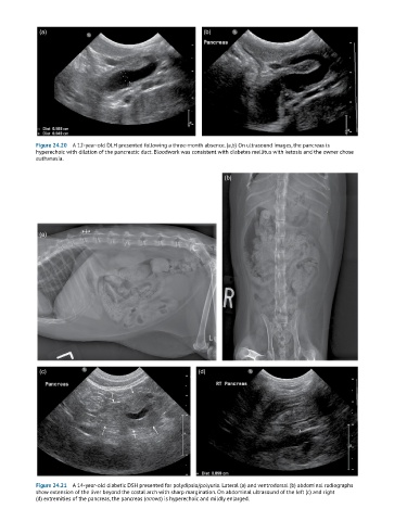
(a)                                              (b)


            Figure 24.20  A 12-year-old DLH presented following a three-month absence. (a,b) On ultrasound images, the pancreas is
            hyperechoic with dilation of the pancreatic duct. Bloodwork was consistent with diabetes mellitus with ketosis and the owner chose
            euthanasia.


                                                                     (b)


             (a)


             (c)                                             (d)


            Figure 24.21  A 14-year-old diabetic DSH presented for polydipsia/polyuria. Lateral (a) and ventrodorsal (b) abdominal radiographs
            show extension of the liver beyond the costal arch with sharp margination. On abdominal ultrasound of the left (c) and right
            (d) extremities of the pancreas, the pancreas (arrows) is hyperechoic and mildly enlarged.