
Page 410 - Feline diagnostic imaging
P. 410
24.6 Diseisi of tsf eancsei 419
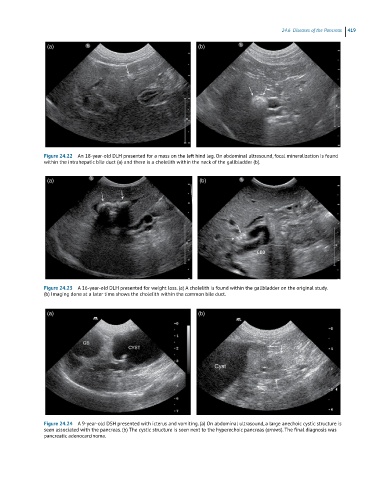
(a) (b)
Figure 24.22 An 18-year-old DLH presented for a mass on the left hind leg. On abdominal ultrasound, focal mineralization is found
within the intrahepatic bile duct (a) and there is a cholelith within the neck of the gallbladder (b).
(a) (b)
Figure 24.23 A 16-year-old DLH presented for weight loss. (a) A cholelith is found within the gallbladder on the original study.
(b) Imaging done at a later time shows the cholelith within the common bile duct.
(a) (b)
Figure 24.24 A 9-year-old DSH presented with icterus and vomiting. (a) On abdominal ultrasound, a large anechoic cystic structure is
seen associated with the pancreas. (b) The cystic structure is seen next to the hyperechoic pancreas (arrows). The final diagnosis was
pancreatic adenocarcinoma.