
Page 415 - Feline diagnostic imaging
P. 415
424 24 Pancreas
Figure 24.35 A female DSH presented with uncontrolled
diabetes and vomiting. The left extremity of the pancreas Figure 24.36 A 15-year-old DSH presented for decreased
(not shown) was enlarged and hypoechoic. In this sonogram, appetite and bloody diarrhea. A large hypoechoic to anechoic
there is a cystic structure (arrows) within the right extremity of nodule is seen in the left extremity of the pancreas. Enlarged
the pancreas. hypoechoic regional lymph nodes and hypoechoic nodules in
the liver increased suspicion for a neoplastic process although
no aspirates were performed.
(a) (b)
Figure 24.37 A 6-year-old DSH with a previous history of pancreatitis diagnosed from surgical biopsy. On presentation, she was
having episodes of vomiting. On ultrasonography, multiple anechoic cystic structures are found associated with the left (a) and right
(b) extremities of the pancreas.
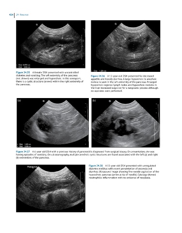
Figure 24.38 A 13-year-old DSH presented with unregulated
diabetes mellitus with recent presentation of anorexia and
diarrhea. Ultrasound image showing fine needle aspiration of the
hypoechoic pancreas (arrow at tip of needle). Cytology showed
neutrophilic inflammation with no evidence of neoplasia.