
Page 412 - Feline diagnostic imaging
P. 412
(a) (b)
(c)
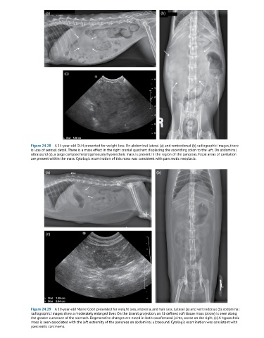
Figure 24.28 A 15-year-old DLH presented for weight loss. On abdominal lateral (a) and ventrodorsal (b) radiographic images, there
is loss of serosal detail. There is a mass effect in the right cranial quadrant displacing the ascending colon to the left. On abdominal
ultrasound (c), a large complex heterogeneously hyperechoic mass is present in the region of the pancreas. Focal areas of cavitation
are present within the mass. Cytologic examination of this mass was consistent with pancreatic neoplasia.
(a) (b)
(c)
Figure 24.29 A 10-year-old Maine Coon presented for weight loss, anorexia, and hair loss. Lateral (a) and ventrodorsal (b) abdominal
radiographic images show a moderately enlarged liver. On the lateral projection, an ill-defined soft tissue mass (arrow) is seen along
the greater curvature of the stomach. Degenerative changes are noted in both coxofemoral joints, worse on the right. (c) A hypoechoic
mass is seen associated with the left extremity of the pancreas on abdominal ultrasound. Cytologic examination was consistent with
pancreatic carcinoma.