
Page 408 - Feline diagnostic imaging
P. 408
24.6 Diseisi of tsf eancsei 417
(a) (b)
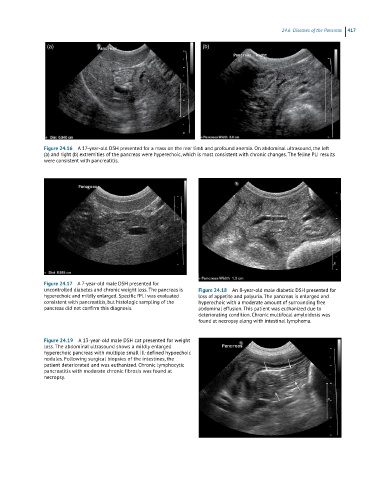
Figure 24.16 A 17-year-old DSH presented for a mass on the rear limb and profound anemia. On abdominal ultrasound, the left
(a) and right (b) extremities of the pancreas were hyperechoic, which is most consistent with chronic changes. The feline PLI results
were consistent with pancreatitis.
Figure 24.17 A 7-year-old male DSH presented for
uncontrolled diabetes and chronic weight loss. The pancreas is Figure 24.18 An 8-year-old male diabetic DSH presented for
hyperechoic and mildly enlarged. Specific fPLI was evaluated loss of appetite and polyuria. The pancreas is enlarged and
consistent with pancreatitis, but histologic sampling of the hyperechoic with a moderate amount of surrounding free
pancreas did not confirm this diagnosis. abdominal effusion. This patient was euthanized due to
deteriorating condition. Chronic multifocal amyloidosis was
found at necropsy along with intestinal lymphoma.
Figure 24.19 A 13-year-old male DSH cat presented for weight
loss. The abdominal ultrasound shows a mildly enlarged
hyperechoic pancreas with multiple small ill-defined hypoechoic
nodules. Following surgical biopsies of the intestines, the
patient deteriorated and was euthanized. Chronic lymphocytic
pancreatitis with moderate chronic fibrosis was found at
necropsy.