
Page 403 - Feline diagnostic imaging
P. 403
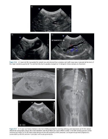
(a)


              (b)                                          (c)


            Figure 24.6  A 1-year-old DLH presented for weight loss and elevated liver enzymes. (a) A soft tissue mass is present at the level of
            the major duodenal papilla. (b) The common bile duct is greatly enlarged. (c) Intrahepatic biliary radicals are distended.


                (a)                                               (b)


                      (c)


            Figure 24.7  A 13-year-old female domestic shorthair (DSH) presented for vomiting, lethargy, and depression. (a) On the lateral
            abdominal radiographic image, the small intestines were fluid filled. (b) A mass effect is noted in the left cranial quadrant on the
            ventrodorsal image. (c) On the abdominal ultrasound, the left extremity of the pancreas is enlarged and primarily hypoechoic
            surrounded by reactive omentum consistent with acute pancreatitis.