
Page 400 - Feline diagnostic imaging
P. 400
24.6 Diseisi of tsf eancsei 409
(a) (b)
(c) (d)
(e) (f)
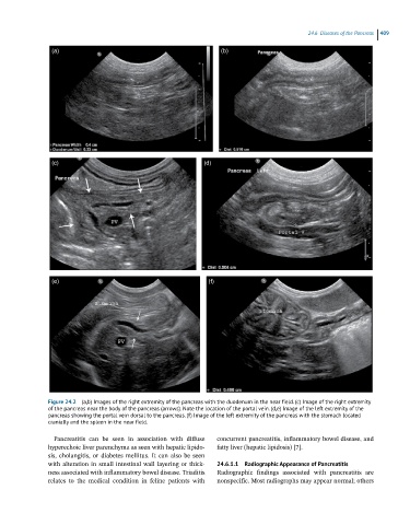
Figure 24.2 (a,b) Images of the right extremity of the pancreas with the duodenum in the near field. (c) Image of the right extremity
of the pancreas near the body of the pancreas (arrows). Note the location of the portal vein. (d,e) Image of the left extremity of the
pancreas showing the portal vein dorsal to the pancreas. (f) Image of the left extremity of the pancreas with the stomach located
cranially and the spleen in the near field.
Pancreatitis can be seen in association with diffuse concurrent pancreatitis, inflammatory bowel disease, and
hyperechoic liver parenchyma as seen with hepatic lipido- fatty liver (hepatic lipidosis) [7].
sis, cholangitis, or diabetes mellitus. It can also be seen
with alteration in small intestinal wall layering or thick- 24.6.1.1 Radiographic Appearance of Pancreatitis
ness associated with inflammatory bowel disease. Triaditis Radiographic findings associated with pancreatitis are
relates to the medical condition in feline patients with nonspecific. Most radiographs may appear normal; others