
Page 401 - Feline diagnostic imaging
P. 401
410 24 Pancreas
(a) (b)
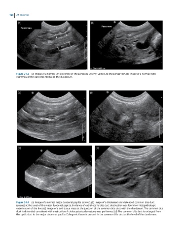
Figure 24.3 (a) Image of a normal left extremity of the pancreas (arrows) ventral to the portal vein. (b) Image of a normal right
extremity of the pancreas medial to the duodenum.
(a) (b)
(c) (d)
Figure 24.4 (a) Image of a normal major duodenal papilla (arrows). (b) Image of a thickened and distended common bile duct
(arrows) at the level of the major duodenal papilla. Evidence of extrahepatic bile duct obstruction was found on histopathologic
examination of the liver. (c) Image of a soft tissue mass at the junction of the common bile duct with the duodenum. The common bile
duct is distended consistent with obstruction. A cholecystoduodenostomy was performed. (d) The common bile duct is enlarged from
the cystic duct to the major duodenal papilla. Echogenic tissue is present in the common bile duct at the level of the duodenum.