
Page 399 - Feline diagnostic imaging
P. 399
408 24 Pancreas
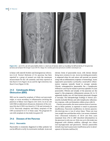
(a) (b)
Figure 24.1 (a) In this cat with pancreatitis, there is a mild loss of serosal detail surrounding the left extremity of the pancreas
(arrows). (b) Compare the image in (a) with this image of a normal pancreas (arrows) in another overweight cat.
is linear with smooth borders and homogeneous echotex- chronic forms of pancreatitis occur, with chronic disease
ture [1,2]. Normal thickness of the pancreas has been being more common in cats. Acute necrotizing pancreatitis
reported in a group of normal cats with the maximum is suspected when fat and acinar cell necrosis are present
measurement for the left extremity and body reported at along with an inflammatory response or hemorrhage. Acute
less than 0.7 cm (Figure 24.3a) and the right extremity less suppurative pancreatitis is primarily a neutrophilic inflam-
than 0.5 cm (Figure 24.3b). matory response typically found in younger animals.
Chronic nonsuppurative pancreatitis has a lymphocytic
infiltration and may be related to previous episodes of acute
24.5 Extrahepatic Biliary pancreatitis. Fibrosis and atrophy of the pancreas are the
Obstruction (EBO) sequel to this progressive inflammatory process [4]. In 31
apparently heathy cats, 45% had chronic pancreatic changes
EBO can be caused by neoplasia of biliary and pancreatic at necropsy. The histopathologic findings of chronic pan-
origin, or occur secondary to inflammation involving the creatitis included fibrosis and mild lymphocytic inflamma-
pancreas or biliary tract (Figures 24.4–24.6). In 21/22 cats tory response, with cyst formation within acinar cells [5].
with EBO on abdominal ultrasound, distension of the com- Chronic pancreatitis, the most common form of pancrea-
mon bile duct (CBD) was found in 17/20 and tortuosity in titis in feline patients, is reported to result in pancreatic
15/19. Pancreatic neoplasia and biliary neoplasia of the insufficiency. Pancreatic insufficiency in chronic pancrea-
CBD accounted for 6/21 of these cases. The prognosis for titis develops due to loss of acinar tissue. These cats may
cats with neoplasia in this study was grave [3]. have weight loss and diarrhea with a low serum cobalamin
level. Ultrasound evaluation of 10/16 cats from cases
24.6 Diseases of the Pancreas examined from 1992 to 2007 described abnormalities in
the pancreas in only one cat. Only one cat in this study was
24.6.1 Pancreatitis diagnosed with diabetes mellitus, although two others
had hyperglycemia and glucosuria. Chronic pancreatitis
Pancreatitis is commonly diagnosed in feline patients but initially may lead to pancreatic insufficiency prior to devel-
can be difficult to verify via noninvasive methods. Acute and opment of diabetes mellitus [6].