
Page 402 - Feline diagnostic imaging
P. 402
24.6 Diseisi of tsf eancsei 411
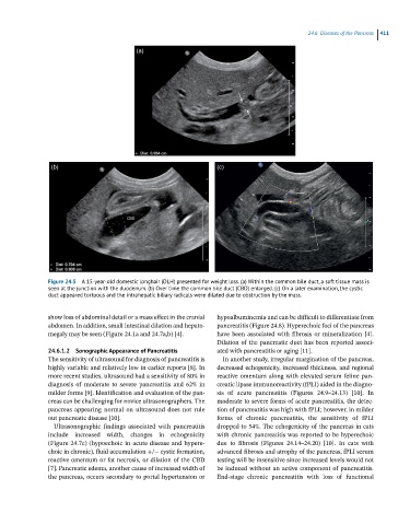
(a)
(b) (c)
Figure 24.5 A 15-year-old domestic longhair (DLH) presented for weight loss. (a) Within the common bile duct, a soft tissue mass is
seen at the junction with the duodenum. (b) Over time the common bile duct (CBD) enlarged. (c) On a later examination, the cystic
duct appeared tortuous and the intrahepatic biliary radicals were dilated due to obstruction by the mass.
show loss of abdominal detail or a mass effect in the cranial hypoalbuminemia and can be difficult to differentiate from
abdomen. In addition, small intestinal dilation and hepato- pancreatitis (Figure 24.8). Hyperechoic foci of the pancreas
megaly may be seen (Figure 24.1a and 24.7a,b) [4]. have been associated with fibrosis or mineralization [4].
Dilation of the pancreatic duct has been reported associ-
24.6.1.2 Sonographic Appearance of Pancreatitis ated with pancreatitis or aging [11].
The sensitivity of ultrasound for diagnosis of pancreatitis is In another study, irregular margination of the pancreas,
highly variable and relatively low in earlier reports [8]. In decreased echogenicity, increased thickness, and regional
more recent studies, ultrasound had a sensitivity of 80% in reactive omentum along with elevated serum feline pan-
diagnosis of moderate to severe pancreatitis and 62% in creatic lipase immunoreactivity (fPLI) aided in the diagno-
milder forms [9]. Identification and evaluation of the pan- sis of acute pancreatitis (Figures 24.9–24.13) [10]. In
creas can be challenging for novice ultrasonographers. The moderate to severe forms of acute pancreatitis, the detec-
pancreas appearing normal on ultrasound does not rule tion of pancreatitis was high with fPLI; however, in milder
out pancreatic disease [10]. forms of chronic pancreatitis, the sensitivity of fPLI
Ultrasonographic findings associated with pancreatitis dropped to 54%. The echogenicity of the pancreas in cats
include increased width, changes in echogenicity with chronic pancreatitis was reported to be hyperechoic
(Figure 24.7c) (hypoechoic in acute disease and hypere- due to fibrosis (Figures 24.14–24.20) [10]. In cats with
choic in chronic), fluid accumulation +/− cystic formation, advanced fibrosis and atrophy of the pancreas, fPLI serum
reactive omentum or fat necrosis, or dilation of the CBD testing will be insensitive since increased levels would not
[7]. Pancreatic edema, another cause of increased width of be induced without an active component of pancreatitis.
the pancreas, occurs secondary to portal hypertension or End‐stage chronic pancreatitis with loss of functional