
Page 420 - Feline diagnostic imaging
P. 420
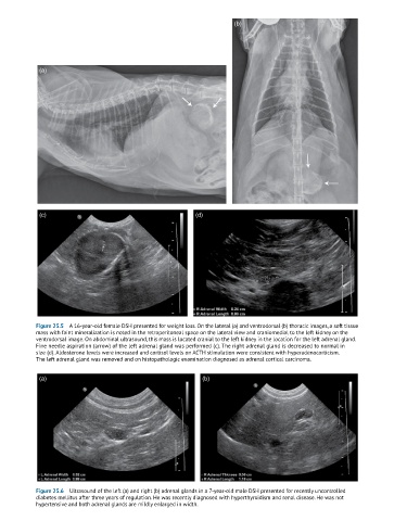
(b)


             (a)


             (c)                                            (d)


            Figure 25.5  A 16-year-old female DSH presented for weight loss. On the lateral (a) and ventrodorsal (b) thoracic images, a soft tissue
            mass with faint mineralization is noted in the retroperitoneal space on the lateral view and craniomedial to the left kidney on the
            ventrodorsal image. On abdominal ultrasound, this mass is located cranial to the left kidney in the location for the left adrenal gland.
            Fine needle aspiration (arrow) of the left adrenal gland was performed (c). The right adrenal gland is decreased to normal in
            size (d). Aldosterone levels were increased and cortisol levels on ACTH stimulation were consistent with hyperadenocorticism.
            The left adrenal gland was removed and on histopathologic examination diagnosed as adrenal cortical carcinoma.

             (a)                                              (b)


            Figure 25.6  Ultrasound of the left (a) and right (b) adrenal glands in a 7-year-old male DSH presented for recently uncontrolled
            diabetes mellitus after three years of regulation. He was recently diagnosed with hyperthyroidism and renal disease. He was not
            hypertensive and both adrenal glands are mildly enlarged in width.