
Page 421 - Feline diagnostic imaging
P. 421
25.7 Hypoadrenocorticism or Addisonns Disease 431
(a)
(b) (c)
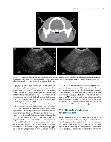
Figure 25.7 An 8-year-old DSH presented for uncontrolled diabetes mellitus. On the postcontrast transverse computed tomographic
image of the brain, there is mild enlargement of the pituitary glands in width (a). Abdominal ultrasound shows enlargement of both
adrenal glands with the right (b) slightly larger than the left (c).
polymyopathy from hypokalemia [4]. Primary HA can one). The size of the affected adrenal glands ranged in thick-
arise from a unilateral adenoma or adenocarcinoma of the ness (10–32 mm) with no difference detected between
adrenal gland or bilateral hyperplasia within the adrenal benign and neoplastic lesions [2]. Advanced imaging studies
cortex (Figures 25.8–25.10). It can cause renal dysfunction of the adrenal glands using computed tomography or mag-
and perpetuate chronic renal disease [7]. Secondary hyper- netic resonance imaging (MRI) have been limited. Prior to
aldosteronism can be activated by hypovolemia from con- surgery, an MRI was performed in two cats because ultra-
gestive heart failure, hypoproteinemia, or chronic renal sound examination could not determine if vascular invasion
failure (Figures 25.11–25.13) [4]. had occurred. MRI showed compression of the CVC by the
In one study of primary hyperaldosteronism, 11/13 cats adrenal masses with no obvious invasion [8].
had unilateral adrenal enlargement on abdominal
ultrasound. The majority presented with weakness due to
hypokalemia. Two cats presented with blindness induced 25.7 Hypoadrenocorticism or
by systemic hypertension although nine others were also Addison’s Disease
hypertensive. Histopathologic examination confirmed that
6/13 cats had unilateral adrenal enlargement with the Addison’s disease is due to decreased production of corti-
diagnosis of adenocarcinoma, 7/13 had adenomas (5/7 uni- costeroid hormones by the adrenal glands. Clinical signs
lateral and 2/7 bilateral) [8]. In a more recent retrospective are lethargy, anorexia, weight loss, depression, weakness,
study, six cats were identified with unilateral adrenal dehydration, and hypothermia. This disease has been
enlargement with variable changes in echogenicity (hypo- reported more commonly secondary to trauma, infiltration
echoic in three, hyperechoic in two, and target lesion in due to lymphoma, or iatrogenic adrenocortical atrophy. No