
Page 423 - Feline diagnostic imaging
P. 423
25.8 eoplasia 433
(a)
(b) (c)
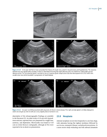
Figure 25.10 A 12-year-old Maine Coon presented for history of weight loss, systemic hypertension, and hypokalemia. The baseline
aldosterone was significantly elevated. A mass is identified involving the right adrenal gland on abdominal radiographs (a) and
ultrasound (b). The left adrenal gland is normal in size (c). A postsurgical complication was the development of a clot within the
caudal vena cava which resulted in progression of renal disease.
(a) (b)
Figure 25.11 A 6-year-old DSH presented with diagnosis of chronic renal disease. The right adrenal gland is mildly enlarged in
width and length (a) and the left (b) is only slightly above normal in size.
description of the ultrasonographic findings are available 25.8 Neoplasia
in the literature [1]. An older study of 10 cats with hypoad-
renocorticism reported that cats presented for weight loss, Adrenal neoplasia occurs less frequently in cats than dogs,
anorexia, and depression. Microcardia was found in 5/10 with adenoma having the highest incidence followed by
cats on initial thoracic radiographs, although all five were carcinoma, metastatic disease, and pheochromocytoma. In
reported to be in shock on presentation. a more recent study evaluating cats with adrenal metastasis