
Page 418 - Feline diagnostic imaging
P. 418
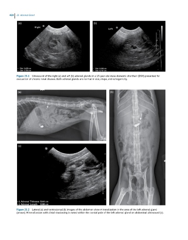
428  25  Adrenal Gland

             (a)                                              (b)


            Figure 25.1  Ultrasound of the right (a) and left (b) adrenal glands in a 13-year-old male domestic shorthair (DSH) presented for
            evaluation of chronic renal disease. Both adrenal glands are normal in size, shape, and echogenicity.


             (a)                                                         (b)


             (c)


            Figure 25.2  Lateral (a) and ventrodorsal (b) images of the abdomen show mineralization in the area of the left adrenal gland
            (arrows). Mineralization with distal shadowing is noted within the cranial pole of the left adrenal gland on abdominal ultrasound (c).