Page 429 - Feline diagnostic imaging
P. 429

440  26  Normal Urinary System

                                                                              (b)


             (a)


            Figure 26.1  Radiographs of the abdomen of a normal cat. (a) Lateral projection showing the right (RK) and left (LK) kidneys. The
            arrow indicates the spleen. L, liver; St, stomach. (b) Ventrodorsal projection. Arrowheads indicate the kidneys. The colon (C) overlies the
            right kidney. The distal extremity of the spleen is superimposed over the left kidney.


                                                              1.9cm


                                                                2.0cm


                                                               2.1cm
                                              5.3cm

                                                             2.3cm
                                                                      5.3cm


                                                                 2.5cm


                                                                2.5cm


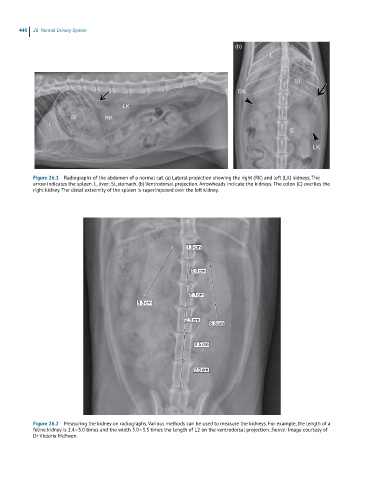
            Figure 26.2  Measuring the kidney on radiographs. Various methods can be used to measure the kidneys. For example, the length of a
            feline kidney is 2.4–3.0 times and the width 3.0–3.5 times the length of L2 on the ventrodorsal projection. Source: Image courtesy of
            Dr Victoria McEwen.
   424   425   426   427   428   429   430   431   432   433   434