
Page 431 - Feline diagnostic imaging
P. 431
442 26 Normal Urinary System
(a) (c)
(b) (d) (e)
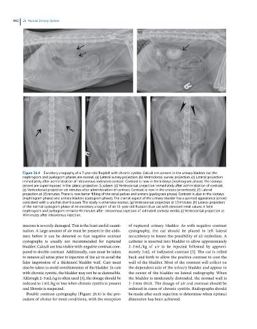
Figure 26.4 Excretory urography of a 7-year-old Ragdoll with chronic cystitis. Calculi are present in the urinary bladder but the
nephrogram and pyelogram phases are normal. (a) Lateral survey projection. (b) Ventrodorsal survey projection. (c) Lateral projection
immediately after administration of intravenous iodinated contrast. Contrast is now in the kidneys (nephrogram phase). The kidneys
(arrow) are superimposed in the lateral projection. S, spleen. (d) Ventrodorsal projection immediately after administration of contrast.
(e) Ventrodorsal projection six minutes after administration of contrast. Contrast is now in the ureters (arrowheads). (f) Lateral
projection at 15 minutes. There is now better filling of the renal pelves and ureters (pyelogram phase). Contrast is also in the kidneys
(nephrogram phase) and urinary bladder (cystogram phase). The cranial aspect of the urinary bladder has a pointed appearance (arrow)
consistent with a urachal diverticulum. The study is otherwise normal. (g) Ventrodorsal projection at 15 minutes. (h) Lateral projection
of the normal cystogram phase of an excretory urogram of an 11-year-old Russian Blue cat with elevated renal values. A faint
nephrogram and pyelogram remains 40 minutes after intravenous injection of iodinated contrast media. (i) Ventrodorsal projection at
40 minutes after intravenous injection.
mucosa is severely damaged. This is the least useful exami- of ruptured urinary bladder. As with negative contrast
nation. A large amount of air must be present in the abdo- cystography, the cat should be placed in left lateral
men before it can be detected so that negative contrast recumbency to lessen the possibility of air embolism. A
cystography is usually not recommended for ruptured catheter is inserted into bladder to allow approximately
bladder. Calculi are less visible with negative contrast com- 2–3 mL/kg of air to be injected followed by approxi-
pared to double contrast. Additionally, care must be taken mately 3 mL of iodinated contrast [3]. The cat is rolled
to remove all urine prior to injection of the air to avoid the back and forth to allow the positive contrast to coat the
false impression of a thickened bladder wall. Care must wall of the bladder. Most of the contrast will collect on
also be taken to avoid overdistension of the bladder. In cats the dependent side of the urinary bladder and appear in
with chronic cystitis, the bladder may not be as distensible. the center of the bladder on lateral radiography. When
Although 2–3 mL/kg is often used [3], the dosage should be the bladder is moderately distended, the normal wall is
reduced to 1 mL/kg or less when chronic cystitis is present 2–3 mm thick. The dosage of air and contrast should be
and fibrosis is suspected. reduced in cases of chronic cystitis. Radiographs should
Double contrast cystography (Figure 26.6) is the pro - be made after each injection to determine when optimal
cedure of choice for most conditions, with the exception distension has been achieved.