
Page 435 - Feline diagnostic imaging
P. 435
446 26 Normal Urinary System
(a)
(c)
(b)
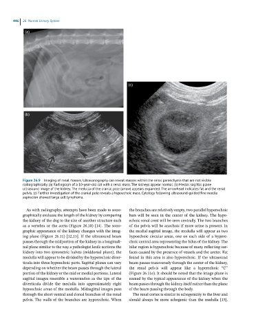
Figure 26.9 Imaging of renal masses. Ultrasonography can reveal masses within the renal parenchyma that are not visible
radiographically. (a) Radiograph of a 10-year-old cat with a renal mass. The kidneys appear normal. (b) Medial sagittal plane
ultrasound image of the kidney. The medulla of the cranial pole (arrow) appears expanded. The arrowhead indicates fat and the renal
pelvis. (c) Further investigation of the cranial pole reveals a hypoechoic mass. Cytology following ultrasound-guided fine needle
aspiration showed large cell lymphoma.
As with radiography, attempts have been made to sono- the branches are relatively empty, two parallel hyperechoic
graphically evaluate the length of the kidney by comparing bars will be seen in the center of the kidney. The hypo-
the kidney of the dog to the size of another structure such echoic renal crest will be seen centrally. The two branches
as a vertebra or the aorta (Figure 26.10) [14]. The sono- of the pelvis will be anechoic if more urine is present. In
graphic appearance of the kidney changes with the imag- the medial sagittal image, the medulla will appear as two
ing plane (Figure 26.11) [12,15]. If the ultrasound beam hypoechoic circular areas, one on each side of a hypere-
passes through the midportion of the kidney in a longitudi- choic central area representing the hilus of the kidney. The
nal plane similar to the way a pathologist knife sections the hilar region is hyperechoic because of many reflecting sur-
kidney into two symmetric halves (middorsal plane), the faces caused by the presence of vessels and the ureter. Fat
medulla will appear to be divided by the hyperechoic diver- found in this area is also hyperechoic. If the ultrasound
ticula into three hypoechoic parts. Sagittal planes can vary beam passes transversely through the center of the kidney,
depending on whether the beam passes through the lateral the renal pelvis will appear like a hyperechoic “C”
portion of the kidney or the mid or medial portions. Lateral (Figure 26.11e). It should be noted that the image plane is
sagittal images resemble a watermelon as the tips of the named by the typical appearance of the kidney when the
diverticula divide the medulla into approximately eight beam passes through the kidney itself rather than the plane
hypoechoic areas of the medulla. Midsagittal images pass of the beam passing through the body.
through the short ventral and dorsal branches of the renal The renal cortex is similar in echogenicity to the liver and
pelvis. The walls of the branches are hyperechoic. When should always be more echogenic than the medulla [15].