
Page 434 - Feline diagnostic imaging
P. 434
26.3 Ultrasonography 445
Figure 26.7 Complications of cystography. A double contrast
cystogram was performed in a young cat, resulting in
extravasation of contrast from the urinary bladder (arrowhead).
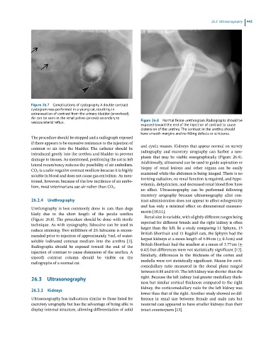
Air can be seen in the renal pelves (arrows) secondary to Figure 26.8 Normal feline urethrogram. Radiographs should be
vesicoureteral reflux.
exposed toward the end of the injection of contrast to cause
distension of the urethra. The contrast in the urethra should
have smooth margins and no filling defects or strictures.
The procedure should be stopped and a radiograph exposed
if there appears to be excessive resistance to the injection of
contrast or air into the bladder. The catheter should be and cystic masses. Kidneys that appear normal on survey
introduced gently into the urethra and bladder to prevent radiography and excretory urography can harbor a neo-
damage to tissues. As mentioned, positioning the cat in left plasm that may be visible sonographically (Figure 26.9).
lateral recumbency reduces the possibility of air embolism. Additionally, ultrasound can be used to guide aspiration or
CO 2 is a safer negative contrast medium because it is highly biopsy of renal lesions and other organs can be easily
soluble in blood and does not cause gas embolism. As men- examined while the abdomen is being imaged. There is no
tioned, however, because of the low incidence of air embo- ionizing radiation, no renal function is required, and hypo-
lism, most veterinarians use air rather than CO 2 . volemia, dehydration, and decreased renal blood flow have
no effect. Ultrasonography can be performed following
excretory urography because ultrasonography after con-
26.2.4 Urethrography trast administration does not appear to affect echogenicity
Urethrography is less commonly done in cats than dogs and has only a minimal effect on dimensional measure-
likely due to the short length of the penile urethra ments [10,11].
(Figure 26.8). The procedure should be done with sterile Renal size is variable, with slightly different ranges being
technique. As with cystography, lidocaine can be used to reported for different breeds and the right kidney is often
reduce straining. Two milliliters of 2% lidocaine is recom- larger than the left. In a study comparing 11 Sphynx, 15
mended prior to injection of approximately 5 mL of water‐ British Shorthair and 15 Ragdoll cats, the Sphynx had the
soluble iodinated contrast medium into the urethra [3]. largest kidneys at a mean length of 4.09 cm (± 0.3 cm) and
Radiographs should be exposed toward the end of the British Shorthair had the smallest at a mean of 3.77 cm (±
injection of contrast to cause distension of the urethra. A 0.43) but differences were not statistically significant [12].
smooth contrast column should be visible on the Similarly, differences in the thickness of the cortex and
radiographs of a normal cat. medulla were not statistically significant. Means for corti-
comedullary ratio measured in the dorsal plane ranged
between 0.88 and 0.93. The left kidney was shorter than the
26.3 Ultrasonography right. Because the left kidney had greater medullary thick-
ness but similar cortical thickness compared to the right
kidney, the corticomedullary ratio for the left kidney was
26.3.1 Kidneys
lower than that of the right. Another study showed no dif-
Ultrasonography has indications similar to those listed for ference in renal size between female and male cats but
excretory urography but has the advantage of being able to neutered cats appeared to have smaller kidneys than their
display internal structure, allowing differentiation of solid intact counterparts [13].