
Page 437 - Feline diagnostic imaging
P. 437
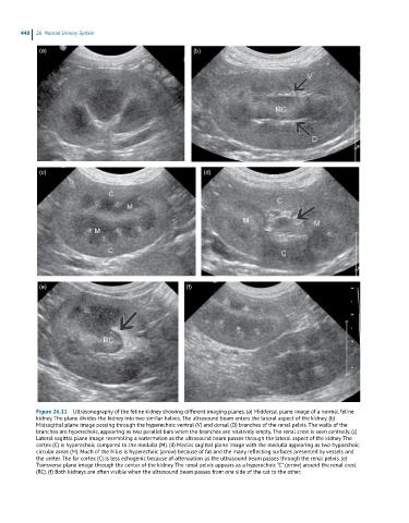
448  26  Normal Urinary System

             (a)                                           (b)


             (c)                                              (d)


             (e)                                         (f)


            Figure 26.11  Ultrasonography of the feline kidney showing different imaging planes. (a) Middorsal plane image of a normal feline
            kidney. The plane divides the kidney into two similar halves. The ultrasound beam enters the lateral aspect of the kidney. (b)
            Midsagittal plane image passing through the hyperechoic ventral (V) and dorsal (D) branches of the renal pelvis. The walls of the
            branches are hyperechoic, appearing as two parallel bars when the branches are relatively empty. The renal crest is seen centrally. (c)
            Lateral sagittal plane image resembling a watermelon as the ultrasound beam passes through the lateral aspect of the kidney. The
            cortex (C) is hyperechoic compared to the medulla (M). (d) Medial sagittal plane image with the medulla appearing as two hypoechoic
            circular areas (M). Much of the hilus is hyperechoic (arrow) because of fat and the many reflecting surfaces presented by vessels and
            the ureter. The far cortex (C) is less echogenic because of attenuation as the ultrasound beam passes through the renal pelvis. (e)
            Transverse plane image through the center of the kidney. The renal pelvis appears as a hyperechoic “C” (arrow) around the renal crest
            (RC). (f) Both kidneys are often visible when the ultrasound beam passes from one side of the cat to the other.