
Page 439 - Feline diagnostic imaging
P. 439
450 26 Normal Urinary System
26.3.2 Urinary Bladder
Ultrasonography has several advantages compared to cys-
tography. Most importantly, most of the risks associated
with cystography can be avoided, including air embolism,
rupture of the urethra or bladder, and iatrogenic infection.
Both procedures can be done if necessary. Previous use of
positive contrast does not affect the quality of ultrasonog-
raphy [3] but negative contrast will degrade the study and
should be removed prior to ultrasonography.
Ultrasonography can be performed with the cat in either
dorsal or lateral recumbency. Lateral recumbency may be
preferred if the cat is fractious, frightened, or distressed.
Regardless of how the patient is initially positioned, posi-
tioning should be changed to check if a visualized lesion is
intraluminal or whether it involves the bladder wall. An
intraluminal structure will fall to the dependent side of the
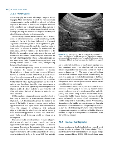
bladder. For example, a mural lesion seen in the near wall Figure 26.14 Ultrasound image of a catheter (white arrow) in
with the cat in left lateral recumbency will be seen on the the urinary bladder of an 11-year-old cat. The catheter appears
far wall when the patient is rotated and placed in right lat- as two parallel hyperechoic lines. The walls (black arrows) of the
eral recumbency. Color Doppler ultrasonography can help bladder are difficult to discern because the bladder is collapsed.
identify vessels within a mural mass, distinguishing
between blood clot and mass. can be uniformly distributed or can form clumps that have
Catheterization is generally avoided since using a cathe- been associated with urine diacylglycerol. Fat should
ter increases the chance of infection or trauma. remain suspended in the lumen and should not result in
Nevertheless, a catheter can be used to control filling of acoustic shadowing [19]. The apex is less well defined
bladder in research or other applications, such as evalua- because of off‐incidence angle artifact. Sound striking the
tion of chemotherapy during drug trials. Sterile gentle pro- apex at an angle can be reflected or refracted so that there
cedure should be used and care should be taken to avoid air appears to be a hole at the apex. Intact tomcats have a dif-
bubble artifacts while injecting saline into the bladder. A fuse prostate that is not easily visualized on ultrasound and
catheter may have been placed for other reasons prior to cannot be seen on radiography.
ultrasound. The wall will appear as parallel echogenic lines In addition to off‐incidence angle artifact, other artifacts
(Figure 26.14). If a Foley catheter is used with the bulb associated with imaging of the urinary bladder include
filled with saline, the bulb will be seen as a circular ane- acoustic enhancement, slice thickness artifact, and side/
choic structure. grating lobe artifact. Deep acoustic enhancement occurs
Mild to moderate bladder distension is preferred so it is because the bladder lumen has few interfaces to reflect
best to try to image the cat before urination has occurred sound and sound is not attenuated as it passes through the
(Figure 26.15). A diuretic can be given if the bladder is too bladder compared to surrounding tissues. Consequently,
empty. If the bladder is too empty, even a normal wall can tissues deep to the bladder are more hyperechoic than adja-
appear thickened. Wall layers can be difficult to visualize cent tissues. Slice thickness and side/grating lobe artifacts
when the bladder is catheterized and collapsed all have the effect of causing structures outside the bladder
(Figure 26.14). On the other hand, the wall of an overly to appear as though they are within the bladder. The most
filled bladder will appear thin or normal even when thick- common example is that adjacent colon can mimic mate-
ened. Early mural thickening could be missed as a rial within the bladder.
consequence.
The normal wall is smooth and has 3–4 layers: a hypere-
choic serosa, a hypoechoic central muscular layer, and a 26.4 Nuclear Scintigraphy
hyperechoic inner layer (Figure 26.15) [18]. The mucosa
may appear hypoechoic. Perivesicular fat is seen around Nuclear scintigraphy has both clinical and research appli-
the apex and neck. The lumen is relatively anechoic but cations. In order to evaluate GFR, Tc99m‐labeled DTPA is
hyperechoic material is frequently suspended in the lumen, injected intravenously and the kidneys are imaged using a
usually attributable to fat in the urine [19]. This material gamma camera (Figure 26.16). GFR is calculated using