
Page 444 - Feline diagnostic imaging
P. 444
(a) (b) (c)
(d) (e) (f)
(g)
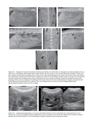
Figure 27.1 Examples of chronic renal disease showing small kidneys and calcification on radiographs. (a) Lateral projection of a cat
with chronic renal disease. Both kidneys have irregular margins and are reduced in size; the left kidney (LK) is smaller than the right
(RK). S, spleen. (b) Ventrodorsal projection of the cat in (a). (c) In this lateral radiograph of a 13-year-old cat with chronic renal disease,
both kidneys are rounded but the right kidney is smaller and contains mineral opacities. (d) Both kidneys are small in this 18-year-old
domestic shorthair with weight loss and chronic renal disease. In particular, the left kidney is small and rounded. (e) Ventrodorsal
projection of the cat in (d). S, spleen. (f) The kidneys of this 6-year-old cat with chronic renal disease are irregularly shaped. The left
one is difficult to locate because of its small size. The cat had hypercalcemia caused by secondary hyperparathyroidism.
(g) Ventrodorsal projection of the cat in (f). S, spleen.
(a) (b)
Figure 27.2 Compensatory hypertrophy in a 14-year-old domestic shorthair. (a) The right kidney was small, measuring 2.41 cm
between the calipers, and misshapen. (b) The right kidney is shown compared to the left. On the scales for each image, the distance
between the horizontal dashes is 1 cm. The left kidney is slightly increased in size but otherwise normal.