
Page 445 - Feline diagnostic imaging
P. 445
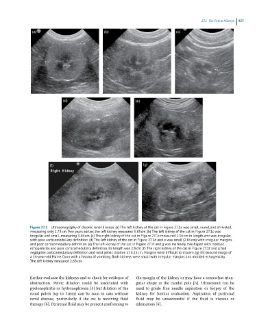
27.1 he eline idneys 457
(a) (b) (c)
(d) (e)
(f) (g)
Figure 27.3 Ultrasonography of chronic renal disease. (a) The left kidney of the cat in Figure 27.1a was small, round, and shriveled,
measuring only 2.73 cm. Two years earlier, the left kidney measured 3.83 cm. (b) The left kidney of the cat in Figure 27.1c was
irregular and small, measuring 3.44 cm. (c) The right kidney of the cat in Figure 27.1c measured 2.16 cm in length and was irregular,
with poor corticomedullary definition. (d) The left kidney of the cat in Figure 27.1d and e was small (2.84 cm) with irregular margins
and poor corticomedullary definition. (e) The left kidney of the cat in Figure 27.1f and g was markedly misshapen with mottled
echogenicity and poor corticomedullary definition. Its length was 2.8 cm. (f) The right kidney of the cat in Figure 27.1f and g had
negligible corticomedullary definition and mild pelvic dilation at 0.23 cm. Margins were difficult to discern. (g) Ultrasound image of
a 16-year-old Maine Coon with a history of vomiting. Both kidneys were small with irregular margins and mottled echogenicity.
The left kidney measured 2.68 cm.
further evaluate the kidneys and to check for evidence of the margin of the kidney or may have a somewhat trian-
obstruction. Pelvic dilation could be associated with gular shape at the caudal pole [6]. Ultrasound can be
pyelonephritis or hydronephrosis [5] but dilation of the used to guide fine needle aspiration or biopsy of the
renal pelvis (up to 3 mm) can be seen in cats without kidney for further evaluation. Aspiration of perirenal
renal disease, particularly if the cat is receiving fluid fluid may be unsuccessful if the fluid is viscous or
therapy [6]. Perirenal fluid may be present conforming to edematous [6].