
Page 450 - Feline diagnostic imaging
P. 450
462 27 Urinary Disease
(a) (b)
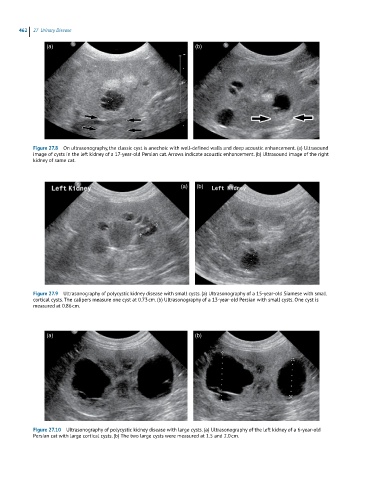
Figure 27.8 On ultrasonography, the classic cyst is anechoic with well-defined walls and deep acoustic enhancement. (a) Ultrasound
image of cysts in the left kidney of a 17-year-old Persian cat. Arrows indicate acoustic enhancement. (b) Ultrasound image of the right
kidney of same cat.
(a) (b)
Figure 27.9 Ultrasonography of polycystic kidney disease with small cysts. (a) Ultrasonography of a 15-year-old Siamese with small
cortical cysts. The calipers measure one cyst at 0.73 cm. (b) Ultrasonography of a 13-year-old Persian with small cysts. One cyst is
measured at 0.86 cm.
(a) (b)
Figure 27.10 Ultrasonography of polycystic kidney disease with large cysts. (a) Ultrasonography of the left kidney of a 6-year-old
Persian cat with large cortical cysts. (b) The two large cysts were measured at 1.5 and 2.0 cm.