
Page 451 - Feline diagnostic imaging
P. 451
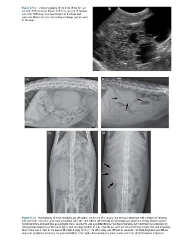
Figure 27.11  Ultrasonography of the liver of the Persian
               cat with PKD shown in Figure 27.8. Five percent of Persian
               cats with PKD may have involvement of the liver and
               pancreas. Numerous cysts including one large cyst are seen
               in the liver.


                  (a)                                               (c)


                                (b)                                 (d)


               Figure 27.12  Radiography of renal neoplasia. (a) Left lateral projection of a 12-year-old domestic shorthair with a history of lethargy
               and vomiting. There is a large mass associated with the right kidney. Differentials include neoplasia, polycystic kidney disease, severe
               hydronephrosis, and perirenal pseudocysts. Renal carcinoma was suspected following ultrasonography but treatment was declined. (b)
               Ventrodorsal projection of the cat in (a). (c) Left lateral projection of a 12-year-old cat with a history of chronic weight loss and bicavitary
               fluid. There was a mass in the area of the right kidney (arrows). The left kidney was difficult to visualize. The final diagnosis was diffuse
               large cell lymphoma involving the gastrointestinal tract, right kidney, mesentery, lymph nodes, and liver. (d) Ventrodorsal projection.