
Page 456 - Feline diagnostic imaging
P. 456
468 27 Urinary Disease
(a) (b)
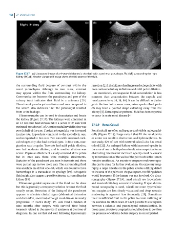
Figure 27.17 (a) Ultrasound image of a 4-year-old domestic shorthair with a perirenal pseudocyst. Fluid (F) surrounding the right
kidney (RK). (b) Another ultrasound image shows the full extent of the fluid.
the surrounding fluid because of contrast within the resection [21], the kidneys had increased echogenicity with
renal parenchyma although in rare cases, contrast poor corticomedullary definition and mild pelvic dilation.
may appear within the fluid surrounding the kidney. As mentioned, extracapsular fluid accumulation is less
Communication between the pseudocyst and part of the common than accumulation between the capsule and
urinary tract indicates that fluid is a urinoma [20]. renal parenchyma [4, 18, 19]. It can be difficult to distin-
Elevation of pseudocyst creatinine and urea compared to guish the two but in some cases, extracapsular fluid pock-
the serum also indicates that the pseudocyst resulted ets may have a pointed shape extending away from the
from urine leakage. kidney [4]. Extracapsular perirenal fluid has been reported
Ultrasonography can be used to characterize and locate to occur in acute renal disease [4].
the kidney (Figure 27.17). The kidneys were abnormal in
all 12 cats that had ultrasound in a series of 26 cats with
perirenal pseudocyst [19]. Corticomedullary definition was 27.1.9 Renal Calculi
poor in half of the cats. Cortical echogenicity was increased Renal calculi are often radiopaque and visible radiographi-
in nine cats, hypoechoic compared to the medulla in one, cally (Figure 27.18). Large calculi that fill the renal pelvis
and unreported in two cats. Two cats with increased corti- or ureter can result in obstruction and hydronephrosis. In
cal echogenicity also had cortical cysts. In four cats, mar- one study, 62% of cats with ureteral calculi also had renal
gination was irregular. Two cats had mild pelvic dilation, calculi [22]. An enlarged kidney with increased opacity in
one had moderate dilation, and in another dilation was the area of one or both pelves should raise suspicion for an
severe. Capsular attachment usually occurred at the pelvis obstructing calculus but increased opacity could be caused
but in three cats, there were multiple attachments. by mineralization of the walls of the pelvis while the lumen
Septation of the pseudocyst was seen in two cats and there remains unaffected. An excretory urogram or ultrasonogra-
were partial tags in two more cats. The surrounding fluid phy can be done for further evaluation. On excretory urog-
was anechoic in all but one cat, which was found to have raphy, a large calculus in the pelvis creates a filling defect
hemorrhage in a transudate on cytology [19]. Echogenic in the area of the pelvis on the pyelogram. No filling defect
fluid might also suggest a possible abscess surrounding the would be present if the lumen was not involved. On ultra-
kidney [18]. sonography (Figure 27.19), renal calculi are hyperechoic
Ultrasound‐guided aspiration of the fluid can be done and most exhibit deep acoustic shadowing. If spatial com-
but this is generally a temporary solution because the fluid pound sonography is used, calculi are more hyperechoic
usually recurs. Resection of the lining of the pseudocyst but margins are less clearly visualized and deep acoustic
appears to alleviate clinical signs (abdominal distension shadowing is apparent less frequently [23]. Sometimes
and discomfort, anorexia) although renal disease is usually there is sufficient fluid in the pelvis to cause outlining of
progressive. In Beck’s study [19], cats lived a median of the calculus. In other cases, it is not possible to distinguish
nine months after surgery with survival time being between a calculus and parenchymal mineralization. In
inversely related to the severity of azotemia at the time of those cases, excretory urography should be done to confirm
diagnosis. In one cat that did well following laparoscopic the presence of calculus before surgery is contemplated.