
Page 457 - Feline diagnostic imaging
P. 457
27.1 he eline idneys 469
(a) (b) (c)
(d) (e) (f)
(g) (h)
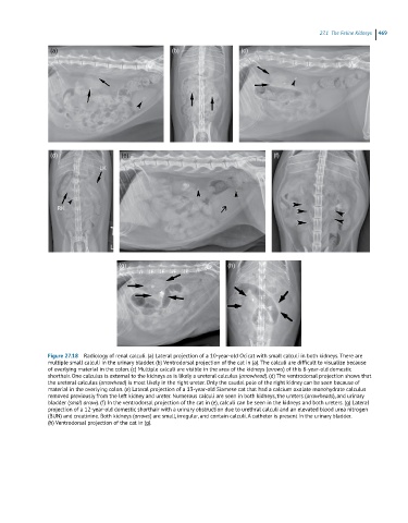
Figure 27.18 Radiology of renal calculi. (a) Lateral projection of a 10-year-old Ocicat with small calculi in both kidneys. There are
multiple small calculi in the urinary bladder. (b) Ventrodorsal projection of the cat in (a). The calculi are difficult to visualize because
of overlying material in the colon. (c) Multiple calculi are visible in the area of the kidneys (arrows) of this 8-year-old domestic
shorthair. One calculus is external to the kidneys as is likely a ureteral calculus (arrowhead). (d) The ventrodorsal projection shows that
the ureteral calculus (arrowhead) is most likely in the right ureter. Only the caudal pole of the right kidney can be seen because of
material in the overlying colon. (e) Lateral projection of a 13-year-old Siamese cat that had a calcium oxalate monohydrate calculus
removed previously from the left kidney and ureter. Numerous calculi are seen in both kidneys, the ureters (arrowheads), and urinary
bladder (small arrow). (f) In the ventrodorsal projection of the cat in (e), calculi can be seen in the kidneys and both ureters. (g) Lateral
projection of a 12-year-old domestic shorthair with a urinary obstruction due to urethral calculi and an elevated blood urea nitrogen
(BUN) and creatinine. Both kidneys (arrows) are small, irregular, and contain calculi. A catheter is present in the urinary bladder.
(h) Ventrodorsal projection of the cat in (g).