
Page 455 - Feline diagnostic imaging
P. 455
27.1 he eline idneys 467
(a) (c) (e)
(b) (d) (f)
(g)
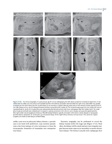
Figure 27.16 Excretory urography of a pseudocyst. (a) On survey radiography, the left lateral projection showed enlargement of one
kidney but the other was very small and rounded. (b) The ventrodorsal projection showed that the right renal silhouette was greatly
enlarged and the left kidney was the small kidney. (c) An excretory urogram (lateral projection) at five minutes shows opacification of
the right kidney with a rim of nonopacification (arrows) surrounding the kidney. (d) The ventrodorsal projection also shows the area of
nonopacification. (e) By 20 minutes, the area surrounding the kidney (arrows) was more radiopaque than the kidney itself. The renal
diverticula (arrowhead) were dilated and increased in opacity consistent with chronic renal disease and possible pyelonephritis.
The right ureter was enlarged and tortuous. The left kidney failed to opacify consistent with chronic renal disease or hypoplasia.
(f) Ventrodorsal projection at 20 minutes. (g) Ultrasound image of the right kidney showing fluid (arrows) in the perirenal region.
Calipers are shown at the margins of the kidney.
unlike cysts seen in polycystic kidney disease, a pseudo- Excretory urography can be performed to reveal the
cyst is not lined with epithelium. Less common pseudo- kidney located within the large cyst (Figure 27.16). Most
cysts arise from leakage of urine (urinomas) or from the often, the kidney will be small and may have irregular mar-
extracapsular formation of transudate into retroperito- gins because most cases occur secondary to severe chronic
neal tissue. renal disease. The kidney is usually more radiopaque than