
Page 454 - Feline diagnostic imaging
P. 454
466 27 Urinary Disease
(a) (d)
(e)
(b)
(c)
(f)
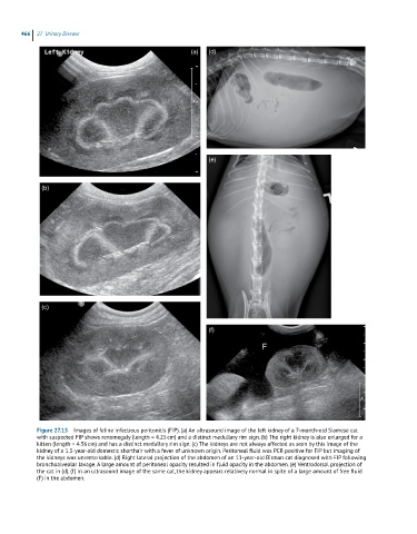
Figure 27.15 Images of feline infectious peritonitis (FIP). (a) An ultrasound image of the left kidney of a 7-month-old Siamese cat
with suspected FIP shows renomegaly (length = 4.25 cm) and a distinct medullary rim sign. (b) The right kidney is also enlarged for a
kitten (length = 4.36 cm) and has a distinct medullary rim sign. (c) The kidneys are not always affected as seen by this image of the
kidney of a 1.5-year-old domestic shorthair with a fever of unknown origin. Peritoneal fluid was PCR positive for FIP but imaging of
the kidneys was unremarkable. (d) Right lateral projection of the abdomen of an 11-year-old Birman cat diagnosed with FIP following
bronchoalveolar lavage. A large amount of peritoneal opacity resulted in fluid opacity in the abdomen. (e) Ventrodorsal projection of
the cat in (d). (f) In an ultrasound image of the same cat, the kidney appears relatively normal in spite of a large amount of free fluid
(F) in the abdomen.