
Page 449 - Feline diagnostic imaging
P. 449
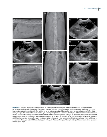
27.1 he eline idneys 461
(b)
(a) (c)
(d) (g)
(f)
(h)
(e)
(i)
Figure 27.7 Imaging of polycystic kidney disease. (a) Lateral projection of a 4-year-old Himalayan cat with polycystic kidneys.
(b) Ventrodorsal projection. Both kidneys are greatly enlarged although the cranial margin of the right kidney is difficult to discern
because the patient is emaciated. (c) The left kidney is markedly enlarged at 5.8 cm and contains large anechoic cysts. (d) The right
kidney is larger at 6.26 cm and contains similar cystic areas. (e) Right lateral projection of an 8-year-old Maine Coon with chronic renal
disease and unilateral polycystic kidney disease. The left kidney is much larger than the right. (f) Ventrodorsal projection of the Maine
Coon showing a normal right kidney and enlarged left kidney. (g) Ultrasound image of the cat in (e) and (f). The renal pelvis is dilated
at 0.79 cm between the calipers. There are multiple small anechoic cysts in the renal cortex. (h) Ultrasound image of the left kidney of
a 6-year-old Himalayan cat with bilateral polycystic kidneys. (i) Ultrasound image of the right kidney of the same cat. Renal cysts can
become quite large.