
Page 446 - Feline diagnostic imaging
P. 446
458 27 Urinary Disease
(a) (b)
(c)
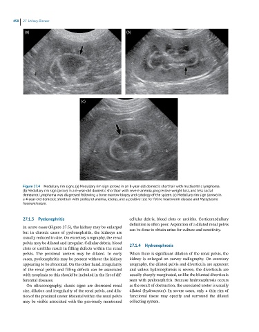
Figure 27.4 Medullary rim signs. (a) Medullary rim sign (arrow) in an 8-year-old domestic shorthair with multicentric lymphoma.
(b) Medullary rim sign (arrow) in a 6-year-old domestic shorthair with severe anemia, progressive weight loss, and less social
demeanor. Lymphoma was diagnosed following a bone marrow biopsy and cytology of the spleen. (c) Medullary rim sign (arrow) in
a 4-year-old domestic shorthair with profound anemia, icterus, and a positive test for feline heartworm disease and Mycoplasma
haemominutum.
27.1.3 Pyelonephritis cellular debris, blood clots or uroliths. Corticomedullary
definition is often poor. Aspiration of a dilated renal pelvis
In acute cases (Figure 27.5), the kidney may be enlarged can be done to obtain urine for culture and sensitivity.
but in chronic cases of pyelonephritis, the kidneys are
usually reduced in size. On excretory urography, the renal
pelvis may be dilated and irregular. Cellular debris, blood 27.1.4 Hydronephrosis
clots or uroliths result in filling defects within the renal
pelvis. The proximal ureters may be dilated. In early When there is significant dilation of the renal pelvis, the
cases, pyelonephritis may be present without the kidney kidney is enlarged on survey radiography. On excretory
appearing to be abnormal. On the other hand, irregularity urography, the dilated pelvis and diverticula are apparent
of the renal pelvis and filling defects can be associated and unless hydronephrosis is severe, the diverticula are
with neoplasia so this should be included in the list of dif- usually sharply marginated, unlike the blunted diverticula
ferential diseases. seen with pyelonephritis. Because hydronephrosis occurs
On ultrasonography, classic signs are decreased renal as the result of obstruction, the associated ureter is usually
size, dilation and irregularity of the renal pelvis, and dila- dilated (hydroureter). In severe cases, only a thin rim of
tion of the proximal ureter. Material within the renal pelvis functional tissue may opacify and surround the dilated
may be visible associated with the previously mentioned collecting system.