
Page 452 - Feline diagnostic imaging
P. 452
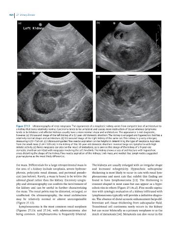
464  27  Urinary Disease

             (a)                                                 (b)


             (c)
                                                                 (d)


            Figure 27.13  Ultrasonography of renal neoplasia. The appearance of a neoplastic kidney varies from complete loss of architecture to
            a kidney that looks relatively normal. Carcinoma tends to be unilateral and causes more destruction of tissue whereas lymphoma
            tends to be bilateral and affected kidneys usually have a more normal shape and architecture. The appearance is not diagnostic,
            however. (a) Ultrasound image of the left kidney of a 12-year-old domestic shorthair. The kidney is enlarged and hyperechoic but has a
            relatively normal shape and architecture. (b) Ultrasound image of the right kidney of the same cat. This kidney is greatly enlarged,
            measuring 6.25 × 7.65 cm. (c) Ultrasound-guided fine needle aspiration can be helpful in determining the type of neoplasia. Aspirates
            from the small mass (1.64 × 1.83 cm) in the kidney of this 10-year-old domestic shorthair revealed large cell lymphoma with high
            mitotic activity. (d) Renal neoplasia can also be the result of metastasis, as is seen in this image of the kidney of a 9-year-old
            domestic shorthair admitted with neoplasia involving the left hindlimb. The kidney shows a loss of architecture with hypoechoic
            areas distorting the shape of the kidney. Fine needle aspiration of the kidneys, limb mass, and medial iliac lymph nodes suggested
            plasmacytoma as the most likely differential.


            the mass. Differentials for a large retroperitoneal mass in   The kidneys are usually enlarged with an irregular shape
            the area of a kidney include neoplasia, severe hydrone-  and  increased  echogenicity.  Hypoechoic  subcapsular
            phrosis, polycystic renal disease, and perirenal pseudo-  thickening is most likely to occur in cats with renal lym-
            cyst (see below). Rarely, a mass is found to be within the   phosarcoma  and  most  cats  that  exhibit  this  finding are
            adrenal gland rather than the kidney. Excretory urogra-  found  to  have  lymphosarcoma  [13].  The  thickening  is
            phy and ultrasonography can confirm the involvement of   crescent shaped in most cases but can appear as a hypo-
            the kidney and can be useful in further characterizing   echoic rim in others (Figure 27.14c,d). Fine needle aspira-
            the mass. The renal pelvis may be distorted, enlarged, or   tion with cytologic evaluation of a kidney infiltrated with
            unaffected.  On  ultrasonography,  the  renal  architecture   lymphosarcoma typically will provide a definitive diagno-
            may  be  relatively  normal  or  almost  unrecognizable   sis. The absence of distal acoustic enhancement helps dif-
            (Figure 27.13).                                   ferentiate soft tissue thickening from subcapsular fluid.
              Lymphosarcoma is the most common renal neoplasm   Transitional  cell  carcinoma  rarely  occurs  in  the  kidney
            (Figures  27.13c  and  27.14),  with  adenocarcinoma  also   but can occur bilaterally as a primary neoplasm or as the
            being  common.  Lymphosarcoma  is  frequently  bilateral.   result of metastasis [14]. Metastasis can also occur in the