
Page 458 - Feline diagnostic imaging
P. 458
470 27 Urinary Disease
(a) (b)
(c) (d)
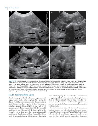
Figure 27.19 Ultrasonography of renal calculi. (a) Ultrasound image of a renal calculus in the left kidney of the cat in Figure 27.18a
and b. The calculus is hyperechoic with deep acoustic shadowing (arrows). (b) Ultrasound image of the right kidney of the cat in
Figure 27.18c and d. The calculus is hyperechoic and exhibits deep acoustic shadowing (arrows). (c) Ultrasound image of the right
kidney of the cat in Figure 21.18e and f. Arrows indicate acoustic shadowing deep to mineralization or calculi in the renal pelvis.
The kidney is misshapen and contains hyperechoic areas consistent with renal infarcts. (d) Ultrasound image of the left kidney of the
cat in Figure 27.18g and h. The kidney is rounded and small with a calculus in the pelvis. Deep acoustic shadowing (arrows) is
apparent. A hyperechoic area likely represents a chronic infarct (I).
27.1.10 Focal Parenchymal Lesions
renal infarction group. The association has been attributed
On ultrasonography, chronic infarcts are hyperechoic tri- to hypercoagulability in these conditions leading to throm-
angular lesions located at the periphery of the renal cortex bosis in vulnerable interlobular arteries, which have no
(Figure 27.20). Indentation may occur as the scar contracts. anastomoses. There was no association with hyperthyroid-
Acute infarcts have been reported to have decreased or ism and unexpectedly, there was a decreased association
mixed echogenicity (Figure 27.20h). In one study of 600 with neoplasia.
cats, concurrent disease was evaluated in 309 cats with Other hyperechoic lesions in the renal parenchyma
renal infarcts and 291 controls [24]. Hypertrophic cardio- include parenchymal mineralization, abscesses, hemor-
myopathy was 4.5 times more likely to occur in cats with rhage, fibrosis, and neoplasia (Figure 27.21). Parenchymal
renal infarcts than in those without the condition. Distal mineralization is consistently hyperechoic and may or
aortic thromboembolism was eight times more likely in the may not have distal acoustic shadowing. Abscesses,