Page 430 - Feline diagnostic imaging
P. 430
26.2 Contrast  adiography  441

                                                                             (b)


                (a)


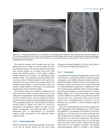
               Figure 26.3  Poorly prepared abdomen. It is important to properly prepare the abdomen before performing an excretory urogram. (a)
               In this lateral projection of a cat with chronic renal disease, one of the kidneys (arrow) is seen to be abnormally small and misshappen.
               (b) In the ventrodorsal projection, neither of the kidneys can be evaluated because of a large amount of fecaloid material in the colon
               superimposed over the kidneys.

                 The  excretory  urogram  can  be  broken  down  into  four     (antegrade ureteropyelography) or into the ureter distal to
               phases that occur in order but overlap (Figure 26.4) [4].   an obstruction (retrograde pyelography) [7].
               The immediate phase is the vascular phase during which
               time  contrast  medium  can  be  found  in  the  aorta,  renal   26.2.3  Cystography
               artery,  and  interlobar  arteries.  As  the  contrast  medium
               reaches  proximal  renal  tubules,  the  nephrogram  phase   Cystography can be performed using positive contrast, neg-
               becomes  visible.  The  nephrogram  should  gradually  fade   ative contrast or a combination of both. In positive contrast
               over the next hour. Contrast medium in the renal pelves,   cystography,  water‐soluble  iodinated  contrast  medium  is
               diverticula, and ureters defines the pyelogram phase. The   delivered through a catheter into the urinary bladder. The
               cystogram  phase  begins  as  contrast  medium  enters  the   iodinated contrast can be either ionic or nonionic although
               bladder. During initiation of this phase, the nephrogram   if a ruptured bladder is suspected in a debilitated cat, non-
               and pyelogram phases will still be visible.        ionic contrast may be preferable. Positive contrast cystogra-
                 The excretory urogram is not without complications but   phy is the best procedure to confirm rupture of the urinary
               these will be fewer when a nonionic rather than an ionic   bladder (Figure 26.5). On the other hand, because small
               iodinated contrast medium is used. Vomiting is common in   calculi are difficult to see within a large amount of positive
               cats but is usually transient. Contrast‐induced renal failure   contrast,  this  is  not  a  good  choice  when  calculi  are  sus-
               is a more serious consequence that is signaled by  persistence   pected. Prior to the administration of contrast, 2–3 mL of
               of the nephrogram phase [6]. Fluid therapy and diuretics   lidocaine  can  be  instilled  to  reduce  straining  during  the
               should  be  given  to  combat  renal  failure  if  it  occurs  [3].   procedure [3].
               Anaphylaxis  and  pulmonary  edema  are  other  rare  but   Room air or carbon dioxide can be used for negative con-
                 serious  consequences.  Care  should  be  taken  to  correct   trast cystography [3,8,9]. Although the use of CO 2  lessens
               dehydration  prior  to  administration  of  intravenous   the possibility of air embolism, the incidence of that com-
                 contrast  media,  particularly  if  ionic  iodinated  contrast   plication is so rare that room air is most commonly used.
               medium is used.                                    The likelihood of air embolism can also be decreased by
                                                                  keeping  the  cat  in  left  lateral  recumbency. This  position
                                                                  will result in any air being more likely to be on the right
               26.2.2  Ureteropyelography
                                                                  side of the heart, where it will be pumped into the lungs
               Instead of iodinated contrast being injected intravenously,   rather  than  into  the  systemic  circulation.  Nevertheless,
               it  can  also  be  injected  directly  into  the  renal  pelvis   pneumocystography  should  be  avoided  if  the  bladder

