
Page 75 - Feline diagnostic imaging
P. 75
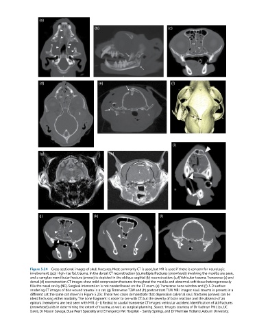
(a)

                                              (b)                                (c)


                    (d)                         (e)                                (f)


                                                                                   (i)

                    (g)                              (h)


                    (j)                       (k)                            (l)


               Figure 5.24  Cross‐sectional images of skull fractures. Most commonly CT is used, but MR is used if there is concern for neurologic
               involvement. (a,b) High‐rise fall trauma. In the dorsal CT reconstruction (a), multiple fractures (arrowheads) involving the maxilla are seen,
               and a complex mandibular fracture (arrows) is depicted in the oblique sagittal (b) reconstruction. (c,d) Vehicular trauma. Transverse (c) and
               dorsal (d) reconstruction CT images show mild compression fractures throughout the maxilla and abnormal soft tissue heterogeneously
               fills the nasal cavity (NC). Surgical intervention is not needed based on the CT exam. (e) Transverse bone window and (f) 3‐D surface
               rendering CT images of bite wound trauma in a cat. (g) Transverse T2W and (h) postcontrast T1W MRI images: skull trauma is present in a
               different cat, the same cat shown in Figure 5.23c. These two cases demonstrate that depression calvarial skull fractures (arrows) can be
               identified using either modality. The bone fragment is easier to see with CT, but the severity of brain reaction and the absence of an
               epidural hematoma are best seen with MRI. (i–l) Rostral to caudal transverse CT images: vehicular accident. Identification of all fractures
               (arrowheads) aids in determining the extent of trauma, as well as surgical planning. Source: Images courtesy of Dr Kathryn Phillips, UC
               Davis, Dr Mason Savage, Blue Pearl Specialty and Emergency Pet Hospital – Sandy Springs, and Dr Merrilee Holland, Auburn University.