
Page 72 - Feline diagnostic imaging
P. 72
68 5 Diagnostic Imaging of Diseases of the Skull
(a) (b)
(c) (d) (e)
(f) (g)
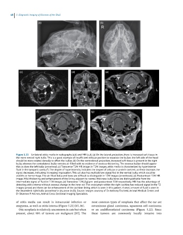
Figure 5.21 Unilateral otitis media in radiographs (a,b) and MRI (c,d). (a) On the lateral projection, there is increased soft tissue in
the more ventral right bulla. This is a good example of insufficient oblique position to separate the bullae; the left side of the head
should be more rotated dorsally to offset the bullae. (b) On the ventrodorsal projection, increased soft tissue is present in the right
bulla, whereas the contralateral bulla remains air filled with no evidence of osseous thickening. The osseous bullae should appear
thin as does the left bulla (arrowhead). (c) Transverse T2W MR image. In T2W images, otitis media is characterized by hyperintense
fluid in the tympanic cavity (*). The degree of hyperintensity indicates the degree of cellular or protein content; as these increase, the
signal decreases, indicating increasing inspissation. This cat also has multiple low signal foci in the ventral bulla, which could be
otoliths or hemorrhage. The air‐filled bulla and bone are difficult to distinguish in T2W images (arrowheads). (d) Postcontrast T1W MR
image. Mild thickening and enhancement of the lining adjacent to normal thickness bulla bone are distinguishable from the
intermediate signal of fluid in T1W images. (e) Transverse T2W, (f,g) pre‐ and postcontrast T1W respectively. MRI has the advantage of
detecting otitis interna without osseous change in the inner ear. The endolymph within the right cochlea has reduced signal in the T2
images (arrow) and there can be enhancement of the cochlear lining, which is seen in this patient. A small amount of fluid is seen in
the dependent right bulla (arrowhead in (e), arrow in (f)). Source: Images courtesy of Dr Anthony Fischetti, Animal Medical Center, and
Dr Shannon P. Holmes, Animal Cross‐Sectional Imaging Specialists.
of otitis media can result in intracranial infection or most common types of neoplasia that affect the ear are
empyema, as well as otitis interna (Figure 5.21) [85, 86]. ceruminous gland carcinoma, squamous cell carcinoma
Otic neoplasia is relatively uncommon in cats but when or an undifferentiated carcinoma (Figure 5.22). Since
present, about 90% of tumors are malignant [87]. The these tumors are commonly locally invasive into