
Page 69 - Feline diagnostic imaging
P. 69
5.1 Diseases of the eline Skull 65
(a) (b) (c)
(d) (e) (f)
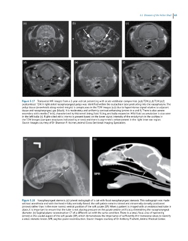
Figure 5.17 Transverse MR images from a 2‐year‐old cat presenting with acute vestibular compromise. (a,b) T2W, (c,d) T1W, (e,f)
postcontrast T1W. A right‐sided nasopharyngeal polyp was identified within the eustachian tube protruding into the nasopharynx. The
polyp tissue (arrowheads along ventral margin) is conspicuous in the T2W images (a,b) due to hyperintense signal relative to adjacent
tissue and nasopharyngeal gas (black). It is moderately and uniformly contrast enhancing (arrows in e and f). There is also severe
secondary otitis media (* in b), characterized by thickened lining, fluid filling, and bulla expansion. Mild fluid accumulation is also seen
in the left bulla (b). Right‐sided otitis interna is present based on the lower signal intensity of the endolymph in the cochlea in
the T2W images (compare structures indicated by arrows) and there is asymmetric enhancement in the right inner ear region.
Source: Images courtesy of Dr Shannon P. Holmes, Animal Cross‐Sectional Imaging Specialists.
(a) (b)
Figure 5.18 Nasopharyngeal stenosis. (a) Lateral radiograph of a cat with focal nasopharyngeal stenosis. This radiograph was made
without anesthesia and with the head mildly ventrally flexed; the soft palate remains kinked and abnormally dorsally positioned
(arrows) rather than in the more normal ventral position of the soft palate (SP). When a patient is imaged with an endotracheal tube in
place, it is important to ensure that the tube is not placing pressure on the palate and/or artificially diminishing the nasopharyngeal
diameter. (b) Sagittal plane reconstruction CT of a different cat with the same condition. There is a small focal area of narrowing
(arrow) at the caudal aspect of the soft palate (SP), which demonstrates the importance of sufficiently thin transverse slices to identify
a small stenotic lesion. SPR, sagittal plane reconstruction. Source: Images courtesy of Dr Anthony Fischetti, Animal Medical Center.