
Page 65 - Feline diagnostic imaging
P. 65
(a) (b) (c)
(d) (e)
(f) (g) (h)
(i) (j)
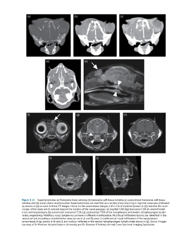
Figure 5.12 Nasal lymphoma. (a) Transverse bone window, (b) transverse soft tissue window, (c) postcontrast transverse soft tissue
window, and (d) dorsal plane reconstruction. Nasal lymphoma can manifest as a solitary mass resulting in regional osteolysis (indicated
by arrows in (a)) as seen in these CT images. Notice on the postcontrast images, a thin rim of contrast (arrows in (d)) outlines the outer
margin of the mass which extends beyond the borders of the nasal passages. (e) Sagittal T2W, (f,g) transverse T2W at conchofrontal
sinus and nasopharynx, (h) precontrast transverse T1W, (i,j) postcontrast T1W of the nasopharynx and medial retropharyngeal lymph
nodes, respectively. Multifocal nasal lymphoma can have a different manifestation. Multifocal infiltrative lesions are identified in the
neurocranium, including a conchofrontal mass (arrow in (e and f)), near circumferential mural infiltration of the nasopharynx
(arrowheads in (g), arrows in (h and i)) and nodular infiltrate in the medial retropharyngeal lymph nodes (arrow in (j)). Source: Images
courtesy of Dr Merrilee Holland, Auburn University, and Dr Shannon P. Holmes, Animal Cross‐Sectional Imaging Specialists.