
Page 61 - Feline diagnostic imaging
P. 61
5.1 Diseases of the eline Skull 57
(a) (b) (c)
(d) (e)
(h)
(f) (g)
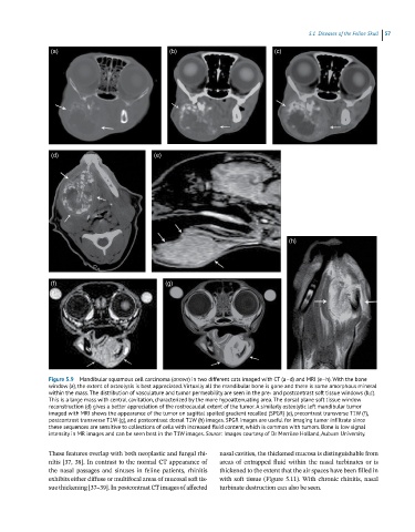
Figure 5.9 Mandibular squamous cell carcinoma (arrows) in two different cats imaged with CT (a–d) and MRI (e–h). With the bone
window (a), the extent of osteolysis is best appreciated. Virtually all the mandibular bone is gone and there is some amorphous mineral
within the mass. The distribution of vasculature and tumor permeability are seen in the pre‐ and postcontrast soft tissue windows (b,c).
This is a large mass with central cavitation, characterized by the more hypoattenuating area. The dorsal plane soft tissue window
reconstruction (d) gives a better appreciation of the rostrocaudal extent of the tumor. A similarly osteolytic left mandibular tumor
imaged with MRI shows the appearance of the tumor on sagittal spoiled gradient recalled (SPGR) (e), precontrast transverse T1W (f),
postcontrast transverse T1W (g), and postcontrast dorsal T1W (h) images. SPGR images are useful for imaging tumor infiltrate since
these sequences are sensitive to collections of cells with increased fluid content, which is common with tumors. Bone is low signal
intensity in MR images and can be seen best in the T1W images. Source: Images courtesy of Dr Merrilee Holland, Auburn University.
These features overlap with both neoplastic and fungal rhi- nasal cavities, the thickened mucosa is distinguishable from
nitis [37, 38]. In contrast to the normal CT appearance of areas of entrapped fluid within the nasal turbinates or is
the nasal passages and sinuses in feline patients, rhinitis thickened to the extent that the air spaces have been filled in
exhibits either diffuse or multifocal areas of mucosal soft tis- with soft tissue (Figure 5.11). With chronic rhinitis, nasal
sue thickening [37–39]. In postcontrast CT images of affected turbinate destruction can also be seen.